Home › Skin discoloration
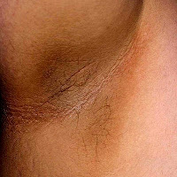
Skin discoloration is the most common dermal condition experienced by us. Though skin discoloration usually may not pose as a problem, in some cases serious diseases may be associated with it.Normally skin coloration and skin pigmentation are dependent on the ethnicity of an individual. On many instances the normal human skin color gets changed in patches or in great areas due to many factors including environment, hormones, foods, immune responses and diseases. The following pages discuss individual skin discolorations and their causes.
Types of skin discoloration
Hypervascularization, inflammation or infections can cause red or pinkish color change in the affected area. Cyanosis, diet, hypercarotenemia, mineral overload, medicines and jaundice can also cause skin discolorations.
For more information read 'Types of skin discoloration'.
Skin discoloration pictures
Patients affected by type 2 diabetes develop scleroderma diabeticorum, a rare disorder of epidermis causing its thickening with excess black or dark brown melanin deposits. Mostly the skin on the upper back and the back of the neck is affected.
For more information read 'Skin discoloration pictures'.
White discoloration on skin
Color changes due to vitiligo are mostly brought about by autoimmune diseases causing death of melanin pigment producing cells (melanocytes). These hypopigmentation patches occur usually on the extremities like fingers. Color changes also occur around body orifices like umbilicus, mouth, genitalia, nostrils and eyes.
For more information read 'White discoloration on skin'.
Dark brown or black discoloration of skin
Melanocytic nevus, commonly known as birthmark, is a common dark brown or black growth of the epidermis. Melanocytic nevus may form subdermally or form as a pigmented growth on the skin. Birthmarks are congenital being present at the time of birth and melanocytic nevi may appear in the later stages of life. Moles and birthmarks which change color, shape or size and those which are painful may have to be medically investigated as some can turn into melanoma (a type of cancer).
For more information read 'Dark brown or black discoloration of skin'.
Reddish skin discoloration
When there is bleeding underneath the dermis purple or reddish discoloration change occurs which is known as purpura. Purpura does not blanch on applying finger pressure while erythema disappears. Inhalation of carbon monoxide can form carboxyhemoglobin in the blood giving reddish coloration. Carbon monoxide inhalation causes debilitating effects at low levels and is fatal in high levels.
For more information read 'Reddish skin discoloration'.
Bluish discoloration of skin
Mongolian spots are due to melanocytes being entrapped and embedded deep in the dermis during their embryonic development and accumulation of melanin. Argyria is the bluish coloration due to accumulation of silver on the dermis caused by ingesting silver compounds as health potions.
For more information read 'Bluish discoloration of skin'.
Yellow discoloration of skin
Increased rate of breakdown of red blood cells can cause pre-hepatic jaundice. Hepatocellular jaundice is caused when the bile does not flow to duodenum. Post-hepatic jaundice is usually due to interruption to the flow of bile inside liver as well as to duodenum.
For more information read 'Yellow discoloration of skin'.
Reddened skin
Nevus flammeus, a birthmark, produces reddened coloration due to dilation of superficial and deeper capillaries. Nevus flammeus usually persists throughout the life. Salmon patches (nevus simplex) are again highly prevalent birthmarks appearing on the forehead, eyelids, knees, on lips or back of neck. Salmon patches are due to dilation of superficial blood vessels and resolve as the child grows.
For more information read 'Reddened skin'.
Orange skin discoloration
The coloration is more pronounced where the stratum corneum is thicker as in palms, soles and nasolabial folds. Secondary carotenemia occurs when there is decreased metabolism or excretion of carotenoids requiring medical treatment. Normally orange coloration resolves over a few days when excess consumption of carrots is stopped.
For more information read 'Orange skin discoloration'.
Related topics:
Homemade moisturizers and treatments.
Tactile sensitivity.
Function of hypodermis.
Skin sensitive to touch.
Methemoglobinemia treatment.
Homemade made face moisturizer recipe.
Methemoglobinemia causes.
Skin senses.
Causes of chronic hives.
Olive oil moisturizer.
Causes of skin sensitivity.
Homemade moisturizers and treatments.
Tactile sensitivity.
Function of hypodermis.
Skin sensitive to touch.
Methemoglobinemia treatment.
Homemade made face moisturizer recipe.
Methemoglobinemia causes.
Skin senses.
Causes of chronic hives.
Olive oil moisturizer.
Causes of skin sensitivity.


No comments:
Post a Comment