Friday, May 31

Skin care - Angioedema definition

Home > Hives and angioedema > Definition of angioedema swellings.
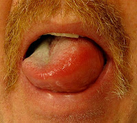
Angioedema is an edema of the subcutaneous tissue usually caused by an allergic reaction to food or drugs. In hives the swelling is superficial skin reactions whereas in angioedema the reaction is under the skin.
The inflammation may range from small spots to large patches of several inches in diameter.

The swellings can occur on any part of the body, but generally occur on the face, around the eyes and lips, inside the mouth and throat and on the tongue. In most of the incidences these reactions resolve by themselves. In some cases when affecting the tongue or throat, the swelling may block the airway and become life threatening.

Angioedema is an immune response to pathogens and triggering factors. It results in production of inflammatory mediators such as histamine by the basophils and mast cells. The resultant inflammatory responses lead to dilation and leakage of blood vessels causing fluid accumulation in the intercellular spaces. In most of the incidences the exact cause may not be known. However many angioedema swelling triggering factors are known. The swelling is usually treated with antihistamine medications.

Medical definitions of angioedema
Wordnetweb.princeton.edu/perl/webwn: "recurrent large circumscribed areas of subcutaneous edema; onset is sudden and it disappears within 24 hours; seen mainly in young women, often as an allergic reaction to food or drugs."

According to thefreedictionary.com: "angioedema, a vascular reaction involving the deep dermis or subcutaneous or submucosal tissues, representing localized edema caused by dilatation and increased permeability of the capillaries, and characterized by the development of giant wheals."
Current topic on skin care:
Angioedema definition.

Wednesday, May 15

Skin care - Hives and angioedema

Home > Angioedema and hives
Hives and angioedema are skin or tissue reactions due to a complex release of inflammatory mediators, including histamine into the surrounding tissues or bloodstream.
In the majority of cases, it is not possible to determinate the exact cause. Many triggering factors of hives and angioedema are known.

Inflammatory mediators such as histamine are produced by basophils and mast cells in the integumentary system and mucosa. Their release is a part of an immune response to pathogens and triggering factors. These inflammatory responses lead to dilation and leakage of blood vessels. The resultant accumulation of body fluids in the intercellular spaces causing the swellings.

Appearance of hives and angioedema

Hives is the result of superficial skin reactions in the upper dermis, leading to pale red, raised bumps. They may cause itchy, stinging or burning sensation. These hives wheals can appear anywhere on the skin surface. The size of the bumps may vary from small spots to large patches of several inches in diameter. Hives may last from a few hours to several weeks.
Angioedema is the result of inflammatory responses leading to rapid swelling of the dermis, subcutaneous tissue, mucosa and submucosal tissues. The angioedema often appears around the eyes, lips, cheeks, tongue and throat. It is also observed to develop on the hands, feet, genitals or intestinal tract. Rapidly progressing angioedema in the mouth and the throat must be treated as a medical emergency as the air passage may get blocked suffocating the patient.

Types and causes hives and angioedema

  • Hives is considered 'idiopathic' if the cause is not known.
  • The skin reaction completely resolving within six weeks is termed 'acute', whereas the condition lasting more than six weeks is known as 'chronic'.
  • Various drugs like dextroamphetamine, penicillin, sulfonamides and anticonvulsants have been found to cause allergic reactions.
  • Exposure to many physical factors such as stress, scratching, firm stroking (dermatographic), pressure, sweating events, exercise (cholinergic), bathing, sun (solar), heat, cold, water (aquagenic), pressure or some types of food can cause hives.
  • Allergens in pollen, animal dander, chemicals, latex, insect venom, cosmetics, detergents etc. can cause skin allergies.
  • Angioedema may occur with or without weals. It is classified as either acquired or hereditary.
  • The acquired skin reactions may be due to unknown causes (idiopathic).
  • The acquired angioedema can be classified as either immunologic or nonimmunologic.
  • It can occur as reactions to hypertension medications like ACE inhibitors.
  • Hereditary angioedema (HAE) is classified into three forms.
  • Hereditary angioedema type I is caused by mutations in the SERPING1 gene, which result in diminished levels of the C1-inhibitor protein.
  • HAE type II is caused by mutations in the SERPING1 gene, which result in the dysfunctional forms of the C1-inhibitor protein.
  • HAE type III is caused by mutations in the F12 gene.
  • All these mutations trigger the complement system causing swellings anywhere in the body.
  • In some persons vibration can cause angioedema, especially of hands.
  • In some patients, angioedema can progress rapidly into Anaphylaxis or anaphylactic shock causing death.

Signs and symptoms hives and angioedema

Symptoms of hives include superficial raised red, pale red or white wheals which can be of varying sizes with itchy sensation. Sometimes there may be burning or stinging sensations. The swellings may last for a few hours to a few days in the acute form and may last for months and years in chronic form.

Angioedema is characterized by large thick swelling. The affected area may be itchy, firm and warm and it may be painful when squeezed or pressed. The swellings may appear on any part of the body. Angioedema may appear as sudden and severe swelling. If it is associated with the gasstrointestinal system, 'abdominal attacks' may occur with symptoms like intense stomach pain and cramps. The swellings in the mouth, tongue, larynx or throat may show symptoms like gasping or wheezy inspiratory breath sounds.
The angioedema may sometime become life-threatening due to blocked airway.

Diagnosis and treatments for hives and angioedema

Mild hives or angioedema does not require treatment. Identifying triggers and avoiding them can help greatly. If the skin reaction is severe, persistent, very itchy or causing severe discomfort, many treatment options are available. Antihistamines are the drugs of choice for treating angioedema and hives.

First-generation antihistamines like hydroxyzine, diphenhydramine and chlorpheniramine are helpful when the symptoms of angioedema are severe and involve swelling on the face, tongue or throat. These medications can cause drowsiness, blurred vision, reduced reflex actions and reduced physical coordination.

Second-generation antihistamines include loratadine, desloratadine, fexofenadine, levocetirizine and cetirizine. These antihistamines have lesser side effects. Chronic cases of hives and angioedema may respond well to  steroid therapy with  prednisone, hydrocortisone, fluocinonide, or desonide. Type H2 antihistamines such as ranitidine, cimetidine and famotidine when given along with type H1 antihistamines treat angioedema and hives.

Epinephrine may be life-saving in severe rapidly progressing allergic angioedema. For treating hereditary angioedema certain androgens, such as dexamethasone, danazol and methyltestosterone have been found to be effective on a long-term basis. In treating hereditary angioedema, Cinryze and Berinert derived from human blood plasma have been found to be effective.
1.Image source: http://en.wikipedia.org/wiki/File:EMminor2010.JPG
2.Image source: http://en.wikipedia.org/wiki/File:Angioedema2013.JPG
Images author: James Heilman, MD
Images license: Creative Commons Attribution-Share Alike 3.0 Unported license.
Current topic on skin care:
Hives and angioedema

Saturday, May 11

Rhinophyma - Causes - Symptoms - Treatment

Home > Symptoms and causes of rhinophyma - treatment of rhinophyma
Rhinophyma is the large, erythemic, bulbous, uneven swelling of the nose caused by the advanced-stage phymatous rosacea. It may affect any person with long-term untreated rosacea.
Rhinophyma is found to occur in middle-aged and elderly men, while it rarely occurs in women. The term rhinophyma is derived from the Greek words, 'rhis' meaning nose and 'phyma' meaning growth, swelling or bulb. It is predominantly found in people of English, Irish and Scottish descent.

The overgrowth (hypertrophy) of sebaceous glands of the tip of the nose can become unsightly and may even obstruct breathing and sight. The effect of rhinophyma disfigurement can leave a strong psychological impact on a patient’s self-esteem leading to depression.

The National Rosacea Society (NRS) has classified rhinophyma as subtype 3 phymatous rosacea. Phymatous rosacea has typical symptoms such as thickening skin, irregular surface nodularities and enlargement. Phymatous rosacea can occur on nose (rhinophyma), forehead (metophyma), ears (otophyma), chin (gnathophyma) and eyelids (blepharophyma).

Causes of rhinophyma

The exact cause and pathogenesis are unknown. The erythema of the nose is due to dilation of the superficial vasculature. The vascularization of the capillaries may cause atrophy of the papillary dermis. The superficial vascularization leads to increased blood flow and causes edema in the nose. Then the overgrowth of sebaceous glands (sebaceous hyperplasia) and fibroplasia follows.

Helicobacter pylori infection is associated with urticaria. Eradication of Helicobacter pylori, improved the phymatous rosacea symptoms. But as there is no total eradication of rosacea, the possibility of H.pylori being the cause remains uncertain.

Alcoholism can cause facial edema and swelling of the nose. But the swelling due to alcoholism or aging is usually even. However alcoholism can worsen the condition. The symptoms of rhinophyma are mistakenly connected to heavy alcohol consumption leading to delay in diagnosis and treatment.

Symptoms presented by rhinophyma

Symptoms of rhinophyma become apparent gradually. Initially there may be intermittent facial flushing. This may be mistaken for age related changes on the skin, sunburn or acne. With the disease progression, symptoms such as erythema and telangiectasias become apparent. This stage is followed by symptoms like skin thickening, fibroplasia and sebaceous hyperplasia of the skin. Typical symptom is the uneven swelling of the nose.

Rhinophyma treatment options

Rhinophyma is a condition that can be easily diagnosed and effectively treated in most patients. If the symptoms are diagnosed early, medication can help in alleviating the condition. Avoiding the triggers and treatment of its earliest manifestations can protect from its progression to later stages and fibrosis.

Medications

Topical metronidazole is the first-line treatment in the early stages. It can be applied in the form of cream or gel once or twice a day. For the patients having tender skin, sulfacetamide lotion may be used as it is less irritating.

Antibiotics like tetracycline, minocycline, doxycycline and clarithromycin have been effective in controlling rhinophyma, when used as simultaneous oral and topical treatments.

Anti-acne medications like topical azelaic acid, retinoic acid and retinaldehyde cream are also effective in controlling the early stages of rhinophyma.

In moderately advanced stages of rhinophyma, the treatment with oral isotretinoin has reduced the sebaceous glands growth and keratinization.

All these medications may have side effects which can be sometime serious. The treatment must be carried out only under the advice of your dermatologist.

Surgical treatment options

The surgery of rhinophyma consists of removing the excess growth. A wide range of surgical procedures is available.

Cryosurgery (cryotherapy) is used to apply extreme cold to destroy the abnormal rhinophyma growth. Freezing temperatures cause ice crystals to form inside the cells, tearing them apart.

Radiofrequency ablation (RFA) is a medical procedure in which a needle-like RFA probe is placed inside the bulbous mass and radiofrequency waves are passing through the probe to increase the temperature within phymatous tissue. It causes destruction of the rhinophyma.

Electrosurgery is a medical procedure in which a high-frequency electric current applied with a needle like electrode to cut, coagulate, desiccate, or fulgurate the rhinophyma tissue.

Tangential excision, CO2 or erbium:YAG laser resurfacing, dermabrasion and skin grafting may be resorted to improve on the final outcome of these surgical rhinophyma treatments.

Rhinophyma image source: http://en.wikipedia.org/wiki/File:Rhinopyma.JPG
Image author: James Heilman, MD.
Image license: Creative Commons Attribution-Share Alike 3.0 Unported license.

Reference:
1.Aaron F. Cohen, MD, and Jeffrey D. Tiemstra, MD. Diagnosis and Treatment of Rosacea. J Am Board Fam Med May 1, 2002 vol. 15 no. 3 214-217.
2.Bolognia JL, Jorizzo JL, Schaffer JV, et al, eds.Dermatology. 3rd ed. Philadelphia, Pa: Mosby Elsevier; 2012:chap 37.
3.Del Rosso JQ, Gallo RL, Kircik L, et al; Why is rosacea considered to be an inflammatory disorder? The primary role, J Drugs Dermatol. 2012 Jun 1;11(6):694-700.

Current topic in skin care:
Causes, symptoms and treatment of rhinophyma.