Thursday, January 31

Epidermal keratinocyte cells in human skin

Home > Know your skin > Epidermis > Keratinocyte cell - Epidermal keratinocytes in human skin
Keratinocytes are epidermal cells found on the outermost layer of the human skin. Nearly 95% of the cells present in the epidermal layer are keratinocytes.
Live and proliferating keratinocytes are found in stratum basale layer. These cells move upwards and differentiate into various layers and finally get sloughed off as dead skin. The epidermal layer is dynamic and is regenerated continuously throughout the human life, turning over every 40-56 days.

Morphogenesis of epidermal keratinocytes

The keratinocytes originate from human epidermal stem cells present in the stratum germinativum (basal layer of epidermis). The progeny of these stem cells are termed transit amplifying (TA) cells. TA cells also reside in the basal layer and undergo mitosis. After a few rounds of mitosis, they undergo differentiation and move outwards. As they move outward, keratinocytes undergo a series of biochemical and morphological changes and pass through various layer-stages. Finally they die and turn into the outermost layer of dead cornecytes (stratum corneum) to be sloughed off.

Structure and function of human keratinocytes

In the basal layer, Keratinocytes are cuboidal or columnar in appearance and are under continuous mitosis. They are separated from dermis by basement membrane and are attached to it through hemidesmosomes. Their function is to continuously divide and proliferate to maintain a regular supply of cells for differentiation in the upper layers.

As the Keratinocytes move upwards into spinosum layer of epidermis, the process of mitosis ceases in them and they undergo keratinization. The nuclei of these cells function actively to synthesize cytokeratin. The cytokeratin, which is a type of fibrillar protein builds up within the keratinocytes and aggregate together to form tonofibrils. Then the tonofibrils form desmosomes. These desmosomes radiate from the keratinocytes and form points of connection between these prickle cells. The net-like structure of these prickle cells provides protection to skin from abrasion and also imparts some elasticity and flexibility to the epidermis. Spinosum keratinocytes produce lamellar bodies having polar lipids, free sterols, phospholipids and enzymes.

As the spinosum keratinocytes are pushed into granulosum layer, the cells have many basophilic granules called keratohyalin granules. Here they are in the process of losing their nuclei and dying. Filaggrin, a type of protein, is found in large quantities in these cells and helps in bundling keratin. In areas of human body like palms and soles where the skin is thick, the lucidum and granulosum layers appear distinct. The differentiation between granulosum and lucidum layers may not be clear where the skin is thin.

As the granulosum keratinocytes are pushed into lucidum layer, they become transparent or translucent and get flattened. The lucidum keratinocytes may be 3-5 layers in thickness and may undergo apoptosis releasing fatty substance. They contain a clear intermediate form of keratin called eleidin. Lucidum keratinocytes are responsible for water-proofing of the epidermis, which is brought about by the release of oily substance.

As the lucidum keratinocytes are moved upward they undergo cornification. Now they are termed as corneocytes. Stratum corneum functions as a barrier protecting the inner layer of epidermis from dehydration and also from invasion of pathogens. Their cell membranes undergo changes and ceramides replace them and a cornified envelope of structural proteins is linked to them. Corneodesmosomes, a type of adhesion proteins, bind the corneocytes. As they move further outward, protein degrading proteases, break them up and the dead skin is sloughed off.
Reference:
1. Maranke I. Koster.(2009). "Making an Epidermis". Annals of the New York Academy of Sciences 1170: 7–10. doi:10.1111/j.1749-6632.2009.04363.x. PMC 2861991. PMID 19686098.
2. Tang, L., Wu, J.J., Ma, Q., Cui, T., Andreopoulos, F.M., Gil, J., Valdes, J., Davis, S.C. and Li, J. (2010), Human lactoferrin stimulates skin keratinocyte function and wound re-epithelialization. British Journal of Dermatology, 163: 38–47. doi: 10.1111/j.1365-2133.2010.09748.x
Current topic: Epidermal keratinocyte cells in human skin.

Get glowing skin complexion. Remove acne scars and blemishes from face.

Wednesday, January 23

Epidermis - Definition of epidermis

Home > Know your skin > Epidermis definition
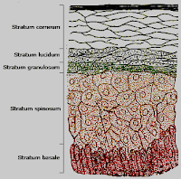
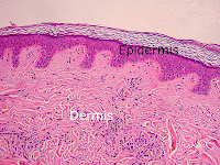
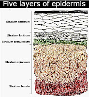
The epidermis is the outermost of the two layers of skin. Epidermis is composed of five well defined layers, namely strata corneum, lucidum, granulosum, spinosum and germinativum.
In structure it is made up of keratinocytes, melanocytes, Langerhans cells and Merkel cells.

The proliferating basal cells differentiate into keratinocytes for forming a natural barrier against the elements and pathogens and also protect the skin from loss of moisture.

Merkel cells present in it are mechanoreceptors and make the skin sensitive to touch. Langerhans cells have immune function and help in eliminating pathogens.
Human skin color complexion is due to the melanin pigment produced by the melanocytes.

Definition of epidermis

As per Wordnetweb.princeton.edu/perl/webwn the definition is:
"the outer layer of the skin covering the exterior body surface of vertebrates."

Thefreedictionary.com's definition is:
"the outer, protective, nonvascular layer of the skin of vertebrates, covering the dermis."

According to Macmillan Dictionary it is:
"the outer layer of skin on top of the dermis. Hair and feathers grow from the epidermis."

According to Merriam-Webster it is:
"the outer epithelial layer of the external integument of the animal body that is derived from the embryonic epiblast; specifically : the outer nonsensitive and nonvascular layer of the skin of a vertebrate that overlies the dermis."

The definition by Oxford Dictionaries is;
"the surface epithelium of the skin of an animal, overlying the dermis."
Reference:
1.Harding, C. R. (2004), The st. corneum: structure and function in health and disease. Dermatologic Therapy, 17: 6–15. doi: 10.1111/j.1396-0296.2004.04S1001.x
Image source:
http://en.wikipedia.org/wiki/File:Skinlayers.png
Author: Serephine
License: Public domain

Current topic: Definition of epidermis.

Get glowing skin complexion. Remove acne scars and blemishes from face.

Monday, January 21

Epidermis parts - Five layers of epidermal skin in order

Home > Know your skin > Epidermis > Descending order of five layers of epidermis
Epidermis is the outermost of the two layers of skin. Epidermal skin is made up of five parts (sublayers).
Its structure is made up of keratinocytes, melanocytes, Langerhans cells and Merkel cells and the majority of them (about 90%) being keratinocytes.

Five layers of epidermis in descending order

Starting from outermost region it is divided into the following five parts or strata in the following order:
  1. Stratum corneum
  2. Stratum lucidum
  3. Stratum granulosum
  4. Stratum spinosum
  5. Stratum basale (also called "stratum germinativum")
Epidermis is avascular (having no blood vessels) and is nourished  by the dermis by diffusion. There is continuous mitotic cell division process going on in keratinocytes in the deepest part of this layer. Each keratinocyte divides into two identical daughter cells of which one remains to carry on the next mitotic cell division and the other one migrates upwards. The older cells are pushed upward and in this process move away from the nourishing dermis. With the loss of nourishment they die and get flattened to form many layers of dead cells, eventually getting sloughed off.

The melanocytes are present in the deepest part and they produce and transfer the skin pigment to the surrounding keratinocytes. Langerhans cells detect the intruding foreign bodies and transport them to lymphocytes present in dermis. Merkel cells present at the deepest part of epidermis function as mechanoreceptors.
Reference:
1.Harding, C. R. (2004), The st. corneum: structure and function in health and disease. Dermatologic Therapy, 17: 6–15. doi: 10.1111/j.1396-0296.2004.04S1001.x
Image source:
http://en.wikipedia.org/wiki/File:Skinlayers.png
Author: Serephine
License: Public domain

Current topic: Parts of epidermis - five layers in descending order.

Get glowing skin complexion. Remove acne scars and blemishes from face.

Sunday, January 20

Stratum granulosum definition - Stratum granulosum layer function

  ›     ›     ›   Definition and function of stratum granulosum layer
Stratum granulosum is a well defined region of epidermis found under the lucidum layer in the palms and soles and under corneum in the other areas of the skin.
As granulosum has visible granules, it is also known as granular layer. Among the five sublayers of epidermis of skin, granulosum is in the middle having spinosum  and lucidum layers on the inner and outer sides.

Definition of stratum granulosum

Stratum granulosum is defined as "the layer of epidermis just under the stratum corneum or (on the palms and soles) just under the stratum lucidum; contains cells (with visible granules) that die and move to the surface." by  wordnetweb.princeton.edu/perl/webwn.

Structure and function of stratum granulosum

In the regions of the body, where the skin is thin, there is no clear cut differentiation between granulosum and lucidum tiers. Under the microscope, this tier appears grainy and consists of 2-5 cell thickness. These grainy, granular or granulosum cells are the keratinocytes, which have migrated from the lower spinosum. These are the last layer of living cells which are in the process of losing their nuclei and dying.

These squamous cells have many basophilic granules called keratohyalin granules which are small and are not bound by membrane. Filaggrin, a type of protein, is found in large quantities in theses cells and is believed to bundle keratin. Lipid containing lamellar granules are present which are bound by membrane. These membrane-bound granules secrete the contents to extracellular space by exocytosis (peni-cytosis) and give rise to sheets of waterproof fatty barrier. This barrier function also prevent nutrients diffusion from the lower cells, leading to death of cells.

Reference:
1.Harding, C. R. (2004), The st. corneum: structure and function in health and disease. Dermatologic Therapy, 17: 6–15. doi: 10.1111/j.1396-0296.2004.04S1001.x
Image source: http://en.wikipedia.org/wiki/File:Skinlayers.png
Author: Serephine
License: Public domain

Current topic in dynamic skin care: Stratum granulosum layer - Definition and function

Get glowing skin complexion. Remove acne scars and blemishes from face.

Tuesday, January 8

Stratum lucidum definition - Function of stratum lucidum layer

  ›     ›     ›   Definition and function of stratum lucidum layer
Stratum lucidum is a well defined epidermal layer found under the corneum region of the skin of the palms and soles. Main function of this stratum is to help the skin handle friction and also waterproof it.
It is a thin transitional layer of epidermis found between the 'horny' and 'granular' layers. However in the rest of the body where the skin is thinner, this stratum does not appear differentiated from the granular region.

Stratum lucidum definition according to Merriam-Webster is: "a thin somewhat translucent layer of cells lying superficial to the stratum granulosum and under the corneum especially in thickened parts of the epidermis (as of the palms or the soles of the feet).
Stratum lucidum is defined as "the layer of epidermis immediately under the corneum in the skin of the palms and soles" by wordnetweb.princeton.edu/perl/webwn
This transparent or translucent layer is composed of three to five tiers of dead keratinocytes. Here these cells do not have distinct boundaries and are flattened. These dead keratinocytes contain a clear intermediate form of keratin called eleidin. The presence of melanosomes may determine the level of its darkness. They are surrounded by fatty substance, probably as the result of the exocytosis of lamellar bodies in the previous tiers. The presence of the oily substance is responsible for the waterproofing function. This barrier function makes the inner regions fairly impermeable to water from outside as well as preventing loss of water from inner cells. Another probable function of this tier is in reduction of friction.

Reference:
1.Harding, C. R. (2004), The st. corneum: structure and function in health and disease. Dermatologic Therapy, 17: 6–15. doi: 10.1111/j.1396-0296.2004.04S1001.x
2.Zirra AM. The functional significance of the skin's st. lcidum. Morphol Embryol (Bucur). 1976 Jan-Mar;22(1):9-12.
1.Image source:
http://en.wikipedia.org/wiki/File:Skinlayers.png
Author: Serephine
License: Public domain
2.Image source:
http://en.wikipedia.org/wiki/File:Epidermal_layers.png
Author: Mikael Häggström, based on work by Wbensmith
License: CC BY-SA 3.0
Current topic in dynamic skin care: Definition and function of stratum lucidum layer

Get glowing skin complexion. Remove acne scars and blemishes from face.

Monday, January 7

Stratum spinosum - Prickle cell layer - Functions of stratum spinosum

  ›     ›     ›   Functions of stratum spinosum (Prickle cell layer)
Stratum spinosum or prickle cell layer of the epidermis has the vital function of Keratinization. Prickle cell layer is the fourth tier from the exterior and is situated between stratum granulosum and germinativum (basale).
The basale and spinosum keratinocytes are together denoted as Malpighian layer. This prickle tier ranges between 5-10 keratinocytes in thickness and the keratinocytes are modified basal keratinocytes having large nuclei which contain very prominent nucleoli.

Keratinization process begins in stratum spinosum. The large pale staining nuclei of prickle layer, function actively to synthesize cytokeratin, a type of fibrillar protein. Cytokeratin builds up within the keratinocytes and aggregate together to form tonofibrils. These tonofibrils form desmosomes which radiate from the keratinocytes forming points of intracellular connections between adjacent prickle keratinocytes. However it is to be noted that there is no continuity of cytoplasm through the 'prickles' of adjacent spinosum keratinocytes.

The name spinosum denotes the prickle or spiny appearance of the keratinocytes due to the radiating desmosomes. This net-like prickle tier provides protection to the underlying tissues. This net-like spinosum also gives some elasticity and flexibility to the epidermis. The prickle cells tier is thicker in areas of skin subject to frequent abrasion such as palms and soles.

One of the important function of the spinosum keratinocytes is the production of lamellar bodies having polar lipids, free sterols, phospholipids and enzymes. Bipolar lipids are produced and retained by these prickle keratinocytes, whose function is prevention of loss of moisture from the skin.

This spinosum layer contains Langerhans cells which are macrophages with the function of protection against pathogens. Langerhans macrophages contain Birbeck granules also known as Birbeck bodies, which are rod shaped with a striated appearance. Birbeck granules function in receptor-mediated endocytosis and phagocytosis. The origin of Langerhans cells is in the bone marrow and they migrate to this prickle region of epidermis. As these polyhedral spinosum keratinocytes approach the granulosum tier they get gradually flattened. Mitosis (cell division function), which occurs continuously in the basale epidermis is nearly absent in these spinosum keratinocytes.

Like the keratinocytes of basale region, spinosum keratinocytes are also susceptible to mutations caused by sunlight and can become malignant. Prickle cell carcinoma is found in fair-skinned individuals who have a history of longer term sun exposure.
Reference:
  • Harding, C. R. (2004), The stratum corneum: structure and function in health and disease. Dermatologic Therapy, 17: 6–15. doi: 10.1111/j.1396-0296.2004.04S1001.x
1.Image source:
http://en.wikipedia.org/wiki/File:Epidermal_layers.png
Author: Mikael Häggström, based on work by Wbensmith
License: CC BY-SA 3.0
2.Image source:
http://en.wikipedia.org/wiki/File:Prickle_cell.JPG
Author: Sandy G Phillips-Long
License: CC BY-SA 3.0

Current topic: Functions of stratum spinosum (Prickle cell layer).

Get glowing skin complexion. Remove acne scars and blemishes from face.

Sunday, January 6

Basal cell layer of epidermal skin - Stratum basale (germinativum)

Home > Know your skin > Epidermis > Basal cell layer of epidermis
Basal cell layer of skin (aka stratum basale or germinativum) is the lowermost sublayer of epidermis. This layer of basal cells is made up of one row of constantly dividing columnar or cubical undifferentiated keratinocytes.
This region is separated from the lower dermis by basement membrane and is attached to the basement membrane through hemidesmosomes.

Structure of basal cell layer

Stratum basale is usually one keratinocyte deep with melanocytes and Langerhans or immune cells dispersed in between. These melanocytes synthesize melanin pigment, giving color and hue to the skin and the hair. The amount of melanin produced though predetermined genetically, is affected by sun exposure levels leading to increase or decrease in production. The Merkel nerve endings (neuroectodermal origin) which have tactile or touch sensation function are also dispersed in this germinativum epidermis. As this basal stratum is in close contact with dermis, it is nurtured with oxygen and nutrients by dermis by diffusion process.

Functions of basal cell layer

High degree of mitosis or cell proliferation activity is found in this basal region and hence the name 'germinativum' (germinating). This cuboid keratinocyte divides into two daughter keratinocytes and the upper keratinocyte migrates towards outer epidermal layers undergoing progressive transformation called keratinisation. After moving into stratum spinosum, these keratinocytes change shape and become polygonal and start synthesizing keratin. Keratinocytes produce more and more keratin and eventually undergo programmed death (apoptosis). In humans, it is estimated that basal stratum turnover from epidermal proliferation units to desquamation every 40–56 days.

The lower basal daughter keratinocyte remains in the stratum basale and undergoes growth and maturation for the next mitosis. As the stratum cornium is continuously sloughed off, the mitosis and replacement activity at basal region should continuously go on. The proliferation and differentiation of basal epidermal region is regulated by Vitamin D3 (cholecalciferol).

Any chemical, physical, physiological, radiological or pathgenic disturbance of this cell proliferation activity at basal germinativum brings about dermal ailments.
Reference:
Harding, C. R. (2004), The stratum corneum: structure and function in health and disease. Dermatologic Therapy, 17: 6–15. doi: 10.1111/j.1396-0296.2004.04S1001.x
Image source:
http://en.wikipedia.org/wiki/File:Epidermal_layers.png
Author:Mikael Häggström, based on work by Wbensmith
License: CC BY-SA 3.0

Current topic: Basal layer cells of epidermal skin.

Get glowing skin complexion. Remove acne scars and blemishes from face.

Saturday, January 5

Stratum corneum layer in skin - Function of stratum corneum cells

Home > Know your skin > Epidermis > Function of the stratum corneum cell layer in the skin
Stratum corneum, the outermost layer of the epidermis, has the skin barrier function and is formed by the death of keratinocyte cells. The stratum corneum (SC) is multi layered and is composed of dead, anucleated, flattened corneocytes.
The stratum corneum has a thickness between 10 and 40 μm and may contain 15-20 layers. These corneocytes are embedded within the multiple lamellar sheets of lipid matrix formed by specialized lipids.

Formation of stratum corneum and desquamation

Epidermis is the outermost layer of skin and it consists of five sublayers. Its main function is to protect underlying tissues and organs from pathogens, toxic agents, oxidants, dehydration, chemicals, environment, ultraviolet radiation and physical stress. Epidermis functions very dynamically and renews continuously.

A continuous process of cell proliferation takes place in the lowermost layer of epidermis known as basale layer. These cells moving outwards undergo maturation and after a number of changes become keratinocyte cells . Eventually these cells die and form the stratum corneum to function as a barrier and get replaced and shed by the desquamation process.

The process of living keratinocytes undergoing transformation into non-living corneocytes is called cornification. As the living keratinocyte cells die, they lose the nuclei and organelles. Their cell membranes undergo changes and ceramides replace them and a cornefied envelope of structural proteins is linked to them.

Desmosomes (corneodesmosomes) which are molecular complexes of cell adhesion proteins and cell linking proteins, bind these corneocytes in the outer st. corneum. As these protein complexes move towards outer region of st. corneum, protein degrading proteases, break them up which leads to desquamation.

Function of stratum corneum

This layer functions as a barrier protecting the inner layer of epidermis from dehydration and also from invasion of pathogens. Stratum corneum contain a dense network of a protein known as keratin. Keratin keeps the epidermis hydrated by absorbing moisture as well as by preventing water evaporation.

The st. corneum by the nature of its structure, makes the epidermis elastic and a glutenous protein bond pulls the epidermis back to its original natural shape.

Another important action of S. corneum is in providing protection from mechanical injuries. To carry out this action, this layer is typically thicker in palms and soles. To some extent it helps in the absorption of moisturizers, holding them back from immediate loss.

Effects of defects in stratum corneum

The formation of corneocytes and their desquamation in a regulated manner is very essential for the maintenance of healthy skin. Any failure or set back in these regulated processes can give rise to skin disorders and ailments. A collection of low-molecular-weight water-soluble components, known as the natural moisturizing factor (NMF) present in st. corneum act as a humectant absorbing moisture by direct contact and also from atmosphere.

These barrier and moisturizing properties can be deranged by intrinsic factors like skin diseases and excessive sebum production or xerosis. Extrinsic factors like weather, temperature, low humidity, surfactants and chemicals can also impair the moisturizing barrier. Mental and physical stress can also have effects on the function of stratum corneum cell layers by the increase in the circulating stress hormones in the skin.
Reference:
Harding, C. R. (2004), The stratum corneum: structure and function in health and disease. Dermatologic Therapy, 17: 6–15. doi: 10.1111/j.1396-0296.2004.04S1001.x
Image source:
http://en.wikipedia.org/wiki/File:Epidermal_layers.png
Author: Mikael Häggström, based on work by Wbensmith
License: CC BY-SA 3.0

Current topic: Function of the stratum corneum layer cells in skin.

Get glowing skin complexion. Remove acne scars and blemishes from face.

Thursday, January 3

Dermis layers - Structure of dermis

Home > Structure of dermis layer
Dermis layer is one of two structural layers of skin. It is located below the epidermis of the skin.
The Dermis, in turn, is made up of upper papillary and lower reticular layers. The structure of this area of skin is made up of a gelatinous ground substance (extrafibrillar and extracellular matrix) containing elastic fibers (yellow fibers), reticular fibers, glycosaminoglycans and collagen. These structures give dermis tensile strength.

Adipocytes (fat cells), fibroblasts, leucocytes and nerve cells are the major types of tissue components found in this region. Depending upon its location on the body, the thickness varies. It is thinnest on the eye lids and thickest on the back. The structure of dermis can also be divided as per its functional tissue components.

Connective tissue
Dense irregular connective tissue, comprising rows of fibroblasts (fiber forming cells) crowded between collagen and elastin fibers, is arranged in sheets in lower region and the upper region is made up of loose connective tissue.

Fat deposits
Fat deposits are found in this region. The thickness of the fat deposits vary in genders and men and women have different areas of fat concentration. Numerous adipocytes (fat cells) are present in groups known as fat lobules.

Vascular tissue
There is a network of blood vessels mainly comprising arterioles, venules and capillaries in this layer.
Extensive network of lymphtic vessels are present in dermis supplyin protective immune cells.

Appendages
Hair follicles are present in this region and are present throughout the body except lips, palms and soles.

Glands
Eccrine glands (sweat glands) are present in the dermis and are dispersed throughout skin regions. Apocrine glands (scent glands) are present in axilla and anogenital regions of dermis. Sebaceous glands (sebum or oil glands) are found in dermis.

Muscles
Smooth muscle structure like Arrector pili muscle is found attached to the base of hair follicle.

Sense organs
There is sensory as well as autonomic nerve network dispersed throughout dermis making skin sensitive to touch and other stimuli. Meissner's corpuscles (touch receptors), Pacinian corpuscles(pressure and vibration receptors), free nerve endings (itch, pain and temperature receptors) and nerve fibers are present.

Immune system
Leukocytes (white blood cells) like neutrophils, monocytes, macrophages, mast cells and T-lymphocytes are present here.
Current topic in skin care: Structure of dermis layer

Get glowing skin complexion. Remove acne scars and blemishes from face.

Wednesday, January 2

Dermis - Papillary layer of dermis

Home > Dermis > Papillary layer of dermis
In dermis, papillary layer lies superficial to the reticular layer. Papillary region lying below the epidermis is closely connecting with it.
It is made up of finger-like (nipple-like) projections, extending into the epidermis. This type of structure strengthens the bond between the skin layers by greatly increasing the surface contact area between these regions.

Papillary region is composed of areolar tissue, the most common type of 'loose connective tissue'. This layer is approximately 300-400 µm deep. It is much thinner when compared to reticular region. Though it is strong enough to bind the skin, it is soft enough to give cushioning and flexibility. It exhibits significant empty space with loosely organised fibers which are interlacing.

Papillary region is dispersed with abundant blood capillaries. Epidermis is avascular (lacking blood vessels). The vasculature of this region supports the epidermis with the supply of nutrients and oxygen for the epidermal cell growth as well as with removal of metabolic wastes. With the unique arrangement for increasing or decreasing the blood flow, thermo-regulatory function of conserving or dissipating body heat is performed by this papillary structure.

The friction ridges (fingerprints) in the palms and fingers and also in the soles and toes are due to the papillary ridges. Apart from giving us grasp, these ridges, with their pattern being controlled genetically, are unique to individuals and provide fingerprints for identity.

Meissner's corpuscles (touch receptors) present in this region in sensitive areas make skin sensitive to touch. One of the important role of papillary layer of dermis is in formation hair, its growth and cycling.
Image source: http://en.wikipedia.org/wiki/File:Normal_Epidermis_and_Dermis_with_Intradermal_Nevus_10x.JPG
Author: Kilbad
License: public domain

Current topic in dynamic skin care: Papillary dermis layer.

Get glowing skin complexion. Remove acne scars and blemishes from face.