Wednesday, December 30

Nevus depigmentosus

Nevus depigmentosus
Nevus depigmentosus is usually a congenital benign non progressive white macule (white patch or spot) with hypopigmentation. Nevus depigmentosus is stable in size and distribution, during the lifetime of the affected individual. The investigation on the histopathology and pathogenesis of this ailment is not complete and its cause is not fully understood.

Symptoms of nevus depigmentosus

These white spots appears as stable spots affecting any part of the body. However they have been found to affect the trunk, hands and feet often.
They do not grow in size. They are usually present from birth and in rare cases they are formed at a later stages of life.
These lesions appear round or oval and grow only with proportion to the growth of the person.
In rare cases certain neurological afflictions have been associated with this ailment; delayed development, seizure disorder and mental retardation have been associated.

Causes of nevus depigmentosus

These white spots are believed to form due to functional abnormalities of the melanocytes and morphological anomalies of melanosomes.
This results in the lowered pigment production in the affected area giving the characteristic pale white spots in the skin.

Differential diagnosis of nevus depigmentosus

Differential diagnosis for this disorder is done to rule out the possibility of affliction by vitiligo (leucoderma) and leprosy.
Unlike leucoderma the white patches or spots are usually present right from birth.
Leucoderma normally show hyperpigmented border to the lesions, whereas in this disorder there is no clear cut hyperpigmented border.
Vitiligo white spots normally grow in size and spread. In rare cases they disappear altogether.

In nevus depigmentosus disorder the white spots are stable and do not spread.
Vitiligo white spots normally occur around the orifices of the body like eyes, ears, nostrils, mouth, umbilicus and genitalia.
Nevus depigmentosus white spots are usually found on the trunk, hands and legs.
The back, chest and buttocks are the most common sites of affliction.

These white spots do not show any alteration of sensitivity or texture of the affected area.
In leprosy the white pale spots occur at a later stage of life and show mild inflammation, hair loss, shine in the skin and numbness. Histological study can differentiate leprosy.
The differential diagnosis for nevus depigmentosus includes hypomelanotic macules of tuberous sclerosis, vitiligo, and anemicus. nevusdepigmentosus
Nevus depigmentosus on the belly

Treatment for nevus depigmentosus

Treatment is not necessary for this ailment. Use of sunscreen and avoiding exposure to sun is advised as the affected area may not pigment on sun exposure; increase in the contrast between the normal skin and the lesion area will affect the aesthetic appearance. There is also the danger of severe sunburn of nevus depigmentosus area on prolonged exposure to sun.

Get glowing skin complexion. Remove acne scars and blemishes from face.

Wednesday, December 23

Skin care - Fordyce spots treatment and cure

White bumps on skin, lips, fingernails, teeth, tonsils and genitaliaFordyce spots overviewFordyce spots treatment and cureNevus depigmentosus
Fordyce spots require no treatment as they are benign and harmless. However for aesthetic appearance, treatment and complete cure is available.

Topical applications for fordyce therapy

The acidic form of vitamin A is Tretinoin and it is also known as ATRA (all-trans retinoic acid).
It is a medicine most commonly used in treating acne.
The topical form is available as gel and cream. For the cure of fordyce spots this ointment must be applied daily till the condition resolves.
Individuals with sensitive skin must use this cautiously and if any adverse reaction starts must discontinue immediately.

Women who are pregnant or who are planning a pregnancy should not use it as tretinoin is a teratogen and can cause fetal abnormalities.
They can only consider the therapy for fordyce spots after child birth.
Better results are achieved when tretinoin used along with AHAs (alpha hydroxy acids) which is another topical solution for acne.
Individuals undergoing therapy for fordyce disease with the above drugs should protect themselves from sun exposure as the treated skin can get severe sun-burn and sun poisoning.
Fordyce's spots on the lips
Fordyce spots on the upper lips (enlarge)

Use of super pulsed laser therapy

For patients seeking improvement of aesthetic appearance from this condition, therapy with CO2 super pulsed laser has been found to be very effective and safe.
Excellent results with complete re-epithelization was achieved.
No side effects were observed and in the medicated area no remnants of the fordyce condition were seen in 15 days.
Diathermy, liquid nitrogen freezing and TCA peels have been used for fordyce spots treatment and cure with varying levels of success.

Get glowing skin complexion. Remove acne scars and blemishes from face.

Saturday, December 19

Milia treatment - milk spots cure - prevent milia

Overview > Causes, signs and symptoms > Milia treatment, cure and how to prevent milia
Milia treatment and cure in newborn babies is to keep the infant clean and well-fed and wait for the baby milk spots to cure by themselves.
Milia are harmless and may take long time to resolve in adults and in some cases may not resolve at all.
Fresh milia milk spots may appear again.
For aesthetic appearance if the affected individual wishes, the milia can removed.
In adults milia treatment, if required, is by exfoliation, dermabrasion, electrodesication, cryosurgery or simply removing the contents by incision and extraction.
Milia treatment and cure by exfoliation
There are a number of exfoliating creams and ointments available.
You can apply once in a day before going to bed.
 In some individuals these creams can cause rash like symptoms and take care to test the suitability of the cream for the treatment by applying it on a small area on your arm.
Alpha Hydroxy Acids containing creams and vitamin A derivative creams have been found to be effective.
Alternately dab and hold a fairly warm towel on the face for 5 minutes to moisten it and make it fairly warm.
This soaks up and softens the milk spots.
Keeping the face moist and warm use a exfoliating scrub to remove surface debris on the skin.
By this treatment the skin covering may loosen and open, causing the contents to come out.
Continue this treatment once in two days till all the milk spots resolve.
The skin on the eyelids is very delicate; take care not to injure it in scrubbing.
Microdermabrasion for the treatment and cure of milia
This is the procedure used in beauty parlours and beauty spas. It is better to have this procedure done by a trained Aesthetician.
Earlier abrasive crystals or wands having rough surface were used. With the advent of technology nowadays a variety of machines are available for microdermabrasion.
Effectiveness varies from person to person and machine to machine. As this procedure is usually priced high think twice before going in for this.
Milia treatment and cure by extraction
It will be better if this is done by a trained aesthetician or a dermatologist.
Face is first cleaned and dabbed with warm face cloth.
With sterile needle the centre of the bump is punctured and a small opening is made.
A gentle squeeze on the spot is made with a suitable forecep (made for such purposes) and the contents are removed. Some antiseptic cream is applied on the cut and it heals in a day or two.
Dermatologists sometime use electrodesication or cryosurgery for the treatment of milk spots.
How to prevent milia
In adults taking certain precautions one can prevent milk spots.
Use only tested skin care products from reputed manufacturers.
Avoid heavy makeup and do not smother the skin with heavy comedogenic lotions and face creams.
To prevent milk spots avoid prolonged sun exposure. If required to go out in the sun, use effective and light sunscreen.
If you are getting milk spots afresh, try periodical exfoliation to prevent milia from forming.
Take medical advice if milk spots are chronic as it could be due to some underlying disease or due to hereditary factor.
Hence for adults milia treatment and cure is possible and by taking precautions it is possible to prevent milia.

Get glowing skin complexion. Remove acne scars and blemishes from face.

Friday, December 18

Milk spots - Baby milk spots - Milia causes

Overview > Milia Causes, signs and symptoms >
Treatment, cure and prevention

Keratin (from dead epidermal cells) causes milia (small epidermoid cysts), when it gets trapped in small pockets on the epidermal surface as cysts/bumps, instead of getting exfoliated (sloughed off) naturally.
The symptoms of milia milk spots are that these small white bumps have no mouth (opening) and on abrasion redden on the edges leaving the central portion white.
Causes of milia in newborn babies
  • This mild skin disorder of baby milk spots or milia show the signs and symptoms as small bead-like raised white bumps on the skin around nose, ears, eyes, cheeks and chin of newborn babies.
  • In some babies these show symptoms like small bumps on the roof of the mouth, gums or genitalia.
  • Milia is very common in newborn babies and its signs and symptoms are found in almost half of them. They usually appear 3-4 days after birth of babies and last for about 20-30 days and resolve naturally.
  • In infants born prematurely the affliction is less and these oil seeds form late (after 2-3 weeks).
  • In rare cases rough bedlinen and clothing has been found to induce the formation of this ailment.
Milk Spots (image by http://www.dermnet.com)
Causes of milia in adults
  • Milk spots can also appear on adult body more often on the face with unknown reasons.
  • Injured or inflamed epidermal region of the body also can form milia in some individuals due to set back in the skin exfoliating process.
  • Among the common and less understood causes of milk spots are the cosmetics and hair care products containing chemicals.
  • Facial creams sometimes smother our skin, preventing sloughing of keratin , dead epidermal cells and sebaceous matter.
  • Comedogenic nature of some facial make-ups, moisturizers and sunscreens (contrary to their claims of being non-comedogenic) causes milia, especially on the eye lids.
  • Some lip balms and lip sticks may be the causes of milk spots on the edge of lips vermilion border.
  • Regular exposure to sun and aging also leads to this skin condition.
  • Certain diseases like PCT (Porphyria Cutanea Tarda ) and epidermolysis bullosa can give rise to these bumps as the secondary disorder.
  • Skin stress and trauma due to radiotherapy and dermabrasion and the use of potent topical corticosteroid, develop symptoms of milia in some persons.
Genetic familial dominant autosomal inheritance disorders have been found to be one of the causes of milia.

Get glowing skin complexion. Remove acne scars and blemishes from face.

Wednesday, December 16

Milk spots - milia - baby milk spots - small white bumps on skin

Causes > Treatment, cure and prevention
Milk spots also called milia, oil seeds or baby milk spots are benign cysts filled with keratin.
They appear as small whitish or slightly pinkish bumps on the skin.
The top most layers of skin die and exfoliate. Milia are actually dead skin trapped as small bumps in pockets on the skin without natural exfoliation.
They also arises in undeveloped sebaceous glands. No opening is visible in it.
They appear usually on the skin around eyes, forehead, cheeks, chin and nose; sometimes they may appear on the genitalia.
Baby milk spots may also appear on gums and the roof of infants mouth (Epstein's pearls).
Baby spots are usually associated with newborns and infants.
About half of the newborn babies develop these. Both boys and girls are affected equally.
In young children baby spots usually disappear within 15-30 days. In infants no treatment is necessary and there are no lasting effects.
However this can affect persons of any age. In older children and adults they may persist and may require removal.
In adults who are very particular about looks, these can be removed.
Removal by an aesthetician or dermatologist is advised.
Secondary oil seeds have been found to occur in persons with sensitive skin stressed by harsh chemicals and heat.
They can also occur on inflamed or injured parts of the body.
They can also arise after a treatment with topical corticosteroids.
Milia (milk spots) on eye lids

It is believed that the milia in some cases have hereditary predisposition and reports of dominant autosomal inheritance of this familial disorder have been documented.

Get glowing skin complexion. Remove acne scars and blemishes from face.

Wednesday, November 25

Tactile defensiveness - Tactile sensitivity - Sensory processing disorder

Tactile defensiveness - tactile sensitivity
Tactile defensiveness (tactile sensitivity) is a sensory processing disorder in which the affected person experiences hyperarousal and exaggerated sensitivity to touch resulting in great distress and discomfort.
Tactile defensiveness is a sensory disorder of the midbrain region wherein the filtering of the incoming stimuli is affected resulting in exaggerated perception of the touch stimuli.
About 75% of the boys with 'fragile X syndrome' and some girls with full X mutation are found to suffer from tactile sensitivity.
The daily life of the tactile defensiveness affected person becomes very miserable. The daily routines like changing dress, brushing teeth and taking bath become arduous tasks. Some of the reactions of the affected children are as follows.
  • The tactile defensiveness affected child may be greatly distressed by gentle touch.He may hate being touched on the face.
  • He may hate coarse clothes. The very feel of the dress on him may distress him.
  • The tactile defensiveness affected person will dislike anything around the neck like tie, jewellery and shirt collar.
  • The person with tactile sensory processing disorder may dislike walking bare-footed and the feel of grass, soil or carpet may annoy him.
  • The tactile defensiveness affected may dislike touching anything slimy and sticky.
  • The sensory processing disorder affected child may dislike brushing teeth and a visit to a dentist becomes an ordeal.
  • The tactile defensiveness affected person may lash out if somebody bumps on to him accidentally or touches or hugs him from behind.
  • Group games are disliked by him and the possibility of touched by others will be avoided by him (even holding hands of partner).
  • He may suffer from bed-wetting, confusion and motor muscle coordination difficulties.
  • Strong preferences of foods may be there and the affected may dislike texture of certain food items.
  • The tactile defensiveness affected person may dislike vibrating toys.
  • He may dislike wind blowing on his body and may so sensitivity to change of temperatures.
The real remedy lies with parents and people in close contact with sensitivity affected child understanding the real problem of tactile defensiveness.
Just imagine a spider crawling on tour arm or your arm being rubbed with a sand paper and your reactions to it.
Similarly a person afflicted with tactile defensiveness has extreme sensitivity and reacts even to normal stimuli.
The sensitivity can be alleviated to great extent by Wilbarger Brushing Protocol, wherein the child's skin is brushed daily with soft surgical brush using a firm pressure.
Repetition of this process over an extent of time will reduce the hyper sensitivity of the skin of the affected person.
The joints of the arms and legs are pressed for sometime regularly. A finger is swiped inside the mouth to desensitize the mouth and reduce sensitivity.
In these treatments there is slow but sure reduction in the tactile defensiveness of the affected child. Soft and seamless clothing for the sensitivity affected children helps a lot.

Current topic: Tactile defensiveness - Tactile sensitivity

Get glowing skin complexion. Remove acne scars and blemishes from face.

Wednesday, November 11

Skin sensitive to touch - tender skin - painful skin

Skin sensitive to touch
Skin becomes sensitive to touch due to a myriad of reasons.
Tender and painful skin condition can be due to its reaction to allergens or underlying adverse health conditions.
There can be two forms of tender conditions to touch. The first being the apparent allergic reactions with lesions and inflammations resulting in tender feeling on touch.
This type of tender condition is dealt in detail in the post "causes of skin sensitivity".
On the other hand there may not be apparent damage or reaction on the skin but it may be touch sensitive, having painful tingling sensation and pinprick sensation.
The cause of this could be due to presence of chemicals and their use in cosmetics, detergents and clothing.
Some foods, food products and many food additives cause tactile hypersensitivity.
Sun exposure and environmental damages can cause the tender and painful feeling.
Hormonal effects and hereditary causes also can cause tactile sensitivity.
Certain health disorders, deficiencies and diseases can also give rise tender feeling to touch.

The deficiencies of thiamine (vitamin B1), biotin (vitamin B7) pyridoxine (vitamin B6) and riboflavin (vitamin B2) have been found to cause hypersensitivity with varied signs like tingling, pin prick and stabbing and painful sensation.
Supplementation with vitamins has been effective in the control of these symptoms.

Diabetes if untreated can cause neuropathy. Diabetic neuropathy affects all peripheral nerves. It can cause skin to be sore and sensitive to touch.
There may be even burning and electric pain.

Migraine apart from being a big bother can also give rise to allodynia (a painful condition of skin being sensitive to touch) and very normal activities like combing can give rise to intense pain.

Herpes zoster is a viral disease and causes blisters with mild to extreme pain with tingling, stinging, aching, stabbing and throbbing sensation.
After the resolution of the attack the lesion area will be very painful for many days to come.

Fibromyalgia (the cause of which is still under debate) can also cause pain and sensitivity of skin to touch.
The affected persons feel widespread painful skin, allodynia (increased response to pressure) and tingling sensation.
The skin may appear normal visually, but becomes highly sensitive to touch.

Peripheral neuropathy is the condition wherein the nerves of the peripheral nervous system are damaged.
There are various causes for this condition. Peripheral neuropathy causes skin to be very sensitive with symptoms like pain, tingling, crawling, itching and pins and needles sensations.
Pain can become very intense.
The affected person becomes so hypersensitive that he cannot stand anything touching certain parts of his body especially his feet.

Demyelineating disorders can give rise to sensitive touch.
There are a number of causes for the occurrence. Myelin sheath around the nerve fibre is destroyed in this ailment.
Paresthesias (sensation of tingling, pricking or stabbing) of the limbs are experienced by the affected persons.

Tactile defensiveness (sensory processing disorder) is a neurological disorder in the the midbrain region.
The stimuli are not properly evaluated and filtered by the brain resulting in exaggerated feeling of touch making the affected person distressed and uncomfortable.
The affected person may not tolerate slightest pressure on the skin.
Sudden appearance of tender feeling to touch can be a hint of progressive damage to the peripheral nerves or even the spinal cord and the brain.

Get glowing skin complexion. Remove acne scars and blemishes from face.

Tuesday, November 10

Skin care - Causes of skin sensitivity

Causes of skin sensitivity

Skin sensitivity

It is a condition wherein a person experiences blown up response of skin to certain stimuli.
A myriad of reactions are experienced by persons with skin sensitivity like, lesions, inflammations, bumps and raised spots, pustules and red and raw spots. They may also experience pain, stinging, numbness and burning sensations on the skin.

Causes of skin sensitivity

Sensitivity of skin is caused by many unknown factors. Among the known factors environment, food, race, age, gender and genetic predisposition play a greater part.
The list of possible causative agents (allergens) and causes is exhaustive. A few of the causes are listed below:
  • Long exposure to sunlight can be a cause
  • Long exposure to heat
  • Long exposure to cold and cold winds
  • High humidity can be a cause
  • Very dry conditions
  • Heated dry air inside the house in winter
  • Salts from sea water and sea-breeze
  • Exposure to radiation and radiotherapy
  • Dust, dust mites and dander of pets
  • Chemicals in cosmetics
  • Chemicals added to food products and preservatives
  • Certain types of foods
  • Flowers and pollen
  • Medicines and formulations
  • Chemotherapy
  • Chemicals in paints and varnish
  • Formalin and formaldehyde present in products around us
  • Pesticides
  • Antibiotics
  • fungicides
  • plastics, polymers, resins, adhesives, lacquers and rubber
  • Contact with metals like, gold, silver, nickel, copper etc...
  • Alkali in the daily-use products
  • Acids in the daily-use products

Manifestations of skin sensitivity

The most common manifestation is eczema or dermatitis. This may lead to itchy lesions which may be dry, scaly and thickened or wet itchy lesions which may be raw and blistered.
  • Chronic photo dermatitis (chronic actinic dermatitis or photo eczema) is a condition wherein dermis reacts to sunlight or artificial light and becomes inflamed.
  • Contact dermatitis, a condition of sensitivity, causes lesion on contact with allergy causing substance (allergen) and is usually localized rash. Mostly surface regions (epidermis) is affected.
    This causes itchy and burning inflammation which may take several days for resolving after the removal of the irritant.
  • Seborrhoeic dermatitis (seborrhoeic eczema) is skin condition caused by unknown factors. This results in red, itchy, scaly and sensitivity and mostly of the scalp and chest are affected.

    Soaps, detergents, hormones and genetic and environmental factors have been found to trigger the rash. In some cases involvement of the fungus Malassezia furfur has been noticed. Excessive intake of vitamin A in children has been associated with this rash. Deficiency of B vitamins like biotin, pyridoxine and riboflavin has been found to cause seborrhoeic dermatitis and sensitivity.
  • Atopic dermatitis is chronic, itchy, relapsing and non-contagious inflammation. Though the exact cause is not known factors like food, chemicals and environment have been found to trigger the rash or worsen the existing condition. The inner side skin of the elbows and knees is the most affected.
  • Migraine affected people have been found to suffer from severe skin pain (cutaneous allodynia). Areas of skin which are subjected slight pressure or scratch are sometimes found to give intense pain in persons with migraine and in some cases scratching may trigger migraine.
Though skin sensitivity cannot be completely prevented, steps can be taken to lessen the situation by mitigating the possible causes. Current topic: Causes of skin sensitivity

Get glowing skin complexion. Remove acne scars and blemishes from face.

Skin senses - human skin senses of pain, touch, cold and warm

Human skin senses

Human skin senses

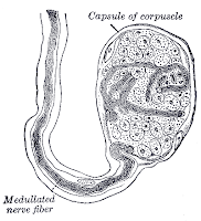
In humans, pain (including itch), cold, warm and touch (including light pressure, deep pressure, vibration and tickle) are perceived by different nerve endings. Skin, the largest and most exposed organ of human body, is endowed with numerous sensory nerve endings.
One square centimeter of human dermis has up to fifty receptor endings for pain, cold, warm and touch. These nerve endings can perceive one or more of senses like touch, pressure, cold, warm and pain. Some areas like tip of the fingers, lips and tongue have more touch receptor endings in human body. There are many types of nerve receptor endings in human skin having specific functions of senses.

Ruffini endings
  • Ruffini corpuscle is a slowly adapting mechanoreceptor existing in the human subcutaneous tissues.
  • This receptor is spindle shaped and perceives the stimulus of stretch and touch.
  • It monitors the slippage along the skin and helps the mechanism of gripping a object.
The Ruffini ending
The Ruffini ending (enlarge)

Meissner's corpuscles
  • Meissner's corpuscles are rapidly adapting mechanoreceptors having highest sensitivity and they convey the senses of light touch.
  • Though they are distributed throughout the human skin, they are more concentrated in fingertips, lips, tongue, soles and palms in human body.
  • They are located primarily just below the epidermis and convey the feeling of touch.
Meissner's corpuscles (sensory nerve ending for touch)
Meissner's corpuscles (sensory nerve ending for touch) (enlarge)

Pacinian corpuscles
  • Pacinian corpuscles are mechanoreceptors in the human skin responsible for pain and pressure senses.
  • This receptors are larger and are fewer in number. The corpuscle about one mm in length and is covered by connective tissue.
  • At the center of the corpuscle is the inner bulb with fluid filled cavity having a single receptor ending without myelin and transmit pain.
Pacinian corpuscles
Pacinian corpuscles (enlarge)

Merkel nerve endings
  • Merkel nerve endings are found in human skin and mucosa providing pressure and texture senses to the brain.
  • The nerve ending consists of Merkel cell and enlarged nerve terminal. These are categorised as slow adapting mechanoreceptors.
Free nerve ending
  • Free nerve ending is unspecialised nerve ending passing on skin pain senses to the human brain.
Thermoreceptors
  • Thermoreceptors are nerve endings in skin conveying the warm and cold perceptions to human brain.
  • There are separate receptors for carrying the warm and cold senses.
  • The warmth receptors are slower in transmitting the impulses when compared to cold receptors.
Nociceptor
  • Nociceptors are receptors that react and convey to human brain the senses of pain. These are specialised pain sensors.
Bulboid corpuscle
  • Bulboid corpuscles are skin receptors which can detect low frequency of vibrations.
    These are minute oval or cylindrical bodies consisting of soft core.
Bulboid corpuscle
Bulboid corpuscle (enlarge)

Hair follicle endings
  • Hair follicle endings respond to displacement of hair and touch senses and are present in hairy areas of the human skin.

Current topic: Human skin senses

Get glowing skin complexion. Remove acne scars and blemishes from face.

Saturday, November 7

Skin care - Olive oil types - Varieties - Extra virgin

NutritionHealth benefitsFor skin careFor hair caremoisturizerBeauty tips
Types of olive oil are many and are classified by International olive oil council depending upon the process of extraction and blending and the aroma, taste, colour and flavour.
Extra virgin olive oil
These types are extracted from the fruits by physical methods and no chemical method is used.
They are the products of first pressing and about 90% of the olive oil is removed at this stage.
To qualify to be called extra virgin, these varieties must not contain more than 0.8% acidity.

Extra virgin types must also have good taste, flavour and aroma.
These varieties are used directly at the table to enhance the flavour of soups, salads and stews and also for dipping.
Premium extra virgin is the finest of the types and contains less than 0.2% acidity.
These types appear yellow to deep green in color and have extra superior flavour with fruity taste.

Virgin olive oil
These types are also produced by the mechanical methods and of the first pressing only.
However these varieties contains extra acidity up to 2%. The rise in acidity when compared to extra virgin varieties may be due to the stage of ripeness of the fruits, the time lapse between the harvest and extraction, the variety of cultivar and the geographic region of the product. It must also have good taste and flavour.
Finer types must contain not more than 1.5% of acidity and should have good flavour.

Refined olive oil
The products which do not qualify to be of higher grades like extra virgin, due to higher and extra acidity are processed further by filtration, heat and chemicals to lower the acidity to 0.3% and remove other defects.
Much of the taste and flavour may be lost. However it is to be noted that solvent extraction method is not involved. This is used in restaurants and in cooking.

Pure olive oil
These types are blends of virgin and refined products.
By addition of virgin olive oil (20%), taste and aroma are infused into the refined product without extra fatty acid percentage (less than 1.5%).
This is priced lesser and hence used in cooking and in restaurants.

immature olives
Immature fruits

Pomace olive oil
By further processing the paste, these lowest types are extracted by treatment with heat and solvents. Blending with virgin olive oil may be done.
But this product is denoted as pomace olive oil. Its acidity should not exceed 1.5%. It is used commercially for making various by-products.


Get glowing skin complexion. Remove acne scars and blemishes from face.

Saturday, October 31

Skin care - Olive oil health benefits

Olive oil typesNutritionHealth benefitsFor skin careFor hair caremoisturizerBeauty tips

Olive oil health benefits are due to the presence of high levels of monounsaturated fatty acids and antioxidants. Prime health benefits of olive oil are in the reduction of risk of heart diseases and certain types of cancers.

Health benefits of olive oil in cardiovascular diseases
Cardiovascular diseases are among the main causes of deaths in the world. These arise out of our lifestyle, food habits, diabetes and use of alcohol and cigarettes.
Premature death are on the rise due to arteriosclerosis (hardening and loss of elasticity of medium and large arteries), atherosclerosis (hardening of arteries due to atheromatous plaques) and coronary heart disease (impaired circulation of blood to cardiac muscles).

The prime cause for these diseases is the presence of low levels of high-density lipoprotein (HDL) cholesterol and high levels of low-density lipoprotein (LDL) cholesterol and triglycerides in the blood.
Cardiovascular disease can be reversed and treated even if the patient has a long history of the ailment.
Reduction in stress and diet control (like replacing fats with olive oil) benefits in preventing, eliminating and reducing almost all forms of heart disease.

Reduction in the overall consumption of fats is the first step in the prevention of heart diseases.
The second step is in replacing whatever fats consumed by monounsaturated fatty acids (MUFA 73% in olive oil).
These fatty acids lower the bad cholesterol (LDL) and increase the good cholesterol (HDL) and thus help in the heart's health.

The wondrous olive oil has a whopping 73% 0f MUFA for our heart's health benefits.
Thus it helps in increasing good cholesterol and lowering bad cholesterol and triglycerides.
Its health benefits in cardiovascular diseases involve in the primary prevention in the development of disease and in the secondary prevention in the recurrence of cardiac event.

Olive oil health benefits as antioxidant
Free radicals are the by-products of our metabolism in our routine activities. They have the capacity to oxidise and damage cells.
Antioxidants are the opposite of free radicals and they help in neutralising the free radicals and give many health benefits.
Vitamin E and phytonutrients like flavonoids and polyphenols present in olive oil have high antioxidant activity and help in protecting our body cells from oxidative damages.

Antioxidants present in olive oil by preventing the oxidation of cholesterol reduce the risk of heart diseases.
Antioxidants have been found to reduce premature ageing and the risk of colon cancer.

Further health benefits of olive oil
Unlike other fats, olive oil can be added and used in our food without cooking and its flavour and taste are appetising.
Olive oil also gives health benefits like lowering blood sugar, preventing peptic ulcers, lowering blood pressure and improving skin condition.
People suffering from ailments like diabetes, gastric ulcers, rheumatic arthritis and asthma have been found to get relief by using olive oil. In skin care olive oil gives immense health benefits like moisturising it and making it soft, smooth, firm and elastic.


Get glowing skin complexion. Remove acne scars and blemishes from face.

Friday, October 30

Skin care - Olive oil nutrition facts

Olive oil typesNutrition factsHealth benefits factsFacts for skin careFor hair careMoisturizerbeauty tips

Exceptional nutrition benefits of olive oil are the presence of high levels of monounsaturated fatty acids, vitamin E and vitamin K.
Olive oil is the main oil ingredient in the food of people of the Mediterranean region. Studies have revealed that heart ailments are comparatively low in this region.

This is attributed to the presence of nearly 73% monounsaturated fatty acids and the antioxidants like vitamin E in the olive extract. Though the exact composition of it varies in respect to extraction process, harvesting time, maturity of the fruits, altitude, region and the genetic line of the trees, the following nutrients are present in approximate values for 100 gms (109 ml).

Olive oil nutrition facts

Nutrients factsAmount in 100gms
Energy890 kcal
Total fats (lipids)100.0 gms
Total saturated fatty acids14.0 gms
Total monounsaturated fatty acids73.0 gms
Total polyunsaturated fatty acids10.5 gms
Phytosterols221.0 mg
Vitamin E (tocopherol)14.4 mg
Vitamin K (phylloquinone)60.0 mcg
Total choline0.3 mg
Betaine0.1 mg
Calcium1.0 mg
Iron0.6 mg
Potassium1.0 mg
Sodium2.0 mg


Facts on the role of fatty acids in olive oil for nutrition and cholesterol management

Saturated fatty acids increase the blood cholesterol levels (both high-density lipoprotein and low-density lipoprotein). The increase in LDL cholesterol leads to artery clogging and heart disease. Monounsaturated fatty acids help in reducing the LDL and increasing HDL. High HDL levels help in removing artery blocks and help in preventing heart disease. Polyunsaturated fatty acids lower both HDL and LDL. The lowering of HDL has adverse effects.

Facts on olive oil role in increasing HDL

The high levels of monounsaturated fatty acids present help in increasing the HDL and lowering the LDL. The facts are that in nutrition remove the deposits on the artery walls allowing better blood flow. The lowering of LDL prevents deposits on the artery walls. Thus olive oil nutrition proves very beneficial for heart care.

Facts on olive oil nutrition increasing antioxidant activity

The high content of vitamin E (a potent antioxidant) and phytonutrients like flavonoids and polyphenols gives a big role in reducing free radical activity. Our body gets protection from oxidative damages which cause many diseases and ageing.

Facts on limitations in olive oil nutrition

The quality of the olive oil is of prime importance. Only 'extra virgin olive oil' is rich in nutrition values. Other label terms like 'pure olive oil', 'refined ' and 'high quality' are misleading terms and facts and the product may not give us the expected benefits. Application for skin nutrition may be avoided by persons affected by tinea versicolor fungal infection as this fungus grows well in it.

Get glowing skin complexion. Remove acne scars and blemishes from face.

Thursday, October 29

Olive oil moisturizer - Homemade skin moisturizer

Homemade olive oil skin moisturizer
Olive oil is an excellent natural skin moisturizer, which can be formulated at home. It is devoid of all side-effects and harms attributed to chemical moisturizer.

What is a moisturizer?

  • Moisturizer is mixture of ingredients designed especially to keep epidermis smooth, soft and elastic.
  • It increases the water content of the epidermis by hydration and reduce loss of water from it by evaporation.
  • Apart from preventing and curing dry conditions, moisturizer also improve texture and tone of the skin.
  • It protects the sensitive and
    tender skin and masks the blemishes and imperfections.
  • They have multiple roles like absorbing water from surroundings and retaining it (humectant) and filling the space between the dermal cells (emollients) and giving smooth texture.

Olive oil as moisturizer

  • Olive oil is obtained by crushing and grinding the fruits. It yields pure extract which is rich in antioxidants.
  • Pure extra virgin olive oil by itself is a good moisturizer.
  • This extra virgin extract on adding to other natural homemade ingredients enhances their hydrating functions.
  • Moisturizers made with extra virgin extract do not normally clog the pores.
  • This is similar to sebum and penetrates the skin exceptionally well.
  • By deep penetration this homemade blend stimulates collagen synthesis making the dermis firm and supple.
  • This homemade blend does not give the greasy feel on application.
  • Homemade moisturizer neutralizes the free radicals in the epidermis and dermis and protects it from ageing and wrinkles.

Homemade olive oil moisturizer

  • Moisturizer blend # 1: Clean your face with a cleanser. Dab it dry with a clean cloth. Do not allow the face to dry out. Within three minutes take 2-3 drops olive oil on your palm and apply it on the face and neck evenly. It retains the moisture and softens the epidermis.
  • Moisturizer blend # 2: If your skin feels a bit dry take equal volumes of oil and water and blend them well. Apply this mix and after ten minutes dab lightly to remove excess mixture. Then apply three drops of the extra virgin extract to lock-in moisture.
  • Olive oil blend # 3: If your face is very dry then try the following method. Add pure honey and olive oil in equal proportion (1/2 teaspoon each) and blend well. Apply this mixture on the face and leave it for 10-15 minutes. Then wash thoroughly with clean water and dab to dry. Immediately apply 2-3 drops of the extra virgin extract to retain moisture. Honey being a good humectant will hold more water in the epidermis and the dryness may disappear within a few days.
  • Moisturizer blend # 4: Alternatively you can take equal volumes of olive and almond oils and blend them well with a little beeswax after heating them. Apply this homemade cream on the face and leave it for 1/2 hour as face mask. Then wash well and apply three drops of it to retain moisture.
  • Blend # 5: If you face has excess sebum, you can blend one tablespoon of olive oil with a few drops of lemon juice, apply and leave it overnight on face. The next day wash well and apply 3 drops of the extra virgin olive extract to retain moisture. This homemade olive oil moisturizer removes only the excess face sebum secretions and does not deplete the skin of its natural secretions.


Current topic:Homemade olive oil skin moisturizer.

Get glowing skin complexion. Remove acne scars and blemishes from face.

Monday, October 26

Pale white spots/patches on children skin face and body

White spots on the face of children

The cause of pale white spots and patches on the face and the body of children must be diagnosed and treated or else it may leave a psychological impact on the kids, apart from the physiological problems.

Various conditions and diseases can cause pale white spots/patches on the skin of children. Some are harmless and resolve by themselves. Many can cause health problems. It is very important for the parents to get the causes diagnosed and have the children treated if necessary.

Pale/white spots and patches arising out of injuries, burns and infections

These white spots are due to skin damage.
Repigmentation of these areas may take a long time or may not develop pigments fully.
These patches can be corrected by chemo or
.

Deficiency of vitamins (avitaminosis) causes pale white spots/patches on the skin

Vitamins are very essential components of our nutrition and the deficiency of them in children can cause many diseases and also affect their skin. Deficiency of B complex vitamins affect our skin in a number of ways.

The deficiency of Vitamin B12 causes pale white spots/patches on the skin especially on the forearms of the kids.These pale patches arise out of depigmentation of the skin and may later turn pink/red causing itching and distress to the kids. Giving food rich in vitamin B12 to the kids makes the pale white vitamin B12 deficiency spots/patches to disappear gradually.

Pale white spots and patches on the children caused by pityriasis alba

This is an asymptomatic condition, the cause of which is not known. The lesions initially appear slightly raised pinkish or reddish in color. Later they flatten out and turn pale white. This condition affects particularly children, more often boys. It usually affects the face of the child, especially cheeks, area around mouth and chin.

Pale white patches may also appear on shoulders arms and hands of kids. By puberty in most of the kids it resolves by itself. The condition may last to a few months or to years. may also recur. No specific treatment is required. Protective and preventive measures may be taken to avoid these pale white spots and patches on your children faces.

Milia (oil seeds or milk spots) causes small raised spots on the face, mouth roof and genitalia

Milia is quite often present in newborns and infants. These white may also affect older children and adults. Milia are keratin filled cysts and are benign and asymptomatic. Usually in newborns they disappear after a few weeks. In adults they may persist and can be removed by opening the cyst.

Fordyce pale white spots

These are usually found on the lips and genitalia. These white spots are actually sebaceous glands which do not have hair follicle association (ectopic sebaceous gland). These are harmless and of cosmetic worry only.

Pale patches caused by nevus depigmentosus

are hypopigmentation areas in the skin and may be present right from the birth. These pale spots are stable, harmless and non- contagious.

Pale white or brown patches/spots caused by tinea (pityriasis) versicolor

Skin yeast and M.globosa cause infections. This ailment usually affects teenagers and young adults. Kids are rarely affected. Malnourished children and kids with suppressed immunity are affected. The fungus can be treated with a number of anti-fungal topical and oral medications.

Pale white spots and patches caused by vitiligo (leucoderma)

is a skin disorder and gives rise to hypopigmentation of some areas of the skin. Autoimmunity is the main . In this ailment the immune cells kill the pigment producing cells (melanocytes) in some areas of the skin leading to white patches.

Pale white spots and patches of leprosy

Leprosy initially shows as pale white spots and patches on the skin, which are numb to touch. Though this is eradicated from most parts of the world, it is still persisting is some pockets of Africa and South east Asia. This is a curable bacterial disease. Areas of skin which lack sensation require immediate medical attention.
Sudden appearance of tender feeling to touch can be a hint of progressive damage to the peripheral nerves or even the spinal cord and the brain.

Pale white spots and patches arising on children must not neglected and immediate diagnosis and treatment must be given.

Get glowing skin complexion. Remove acne scars and blemishes from face.

Sunday, October 18

Pityriasis alba pale/white spots signs symptoms diagnosis

Pityriasis alba > Causes > Diagnosis > Treatment
In the diagnosis of pityriasis alba its signs and symptoms must be analysed to arrive at the differential diagnosis to exclude other conditions and infections.

Signs and symptoms of pityriasis alba
Pityriasis alba is an asymptomatic, self-limiting and benign lesion.
It usually appears on kids of the age 3 -16 years.
The lesions are usually limited to face, especially cheeks, around mouth and chin.
Pityriasis alba may also affect shoulders, neck and arms.
In very rare cases extensive lesions of pityriasis alba may appear all over the body.

Pityriasis alba initially appear like raised red or pink patches sometime with mild itch.
After some time these pityriasis alba patches flatten out and look pale with depigmentation, which is not total.
The spots are oval or round, about 2cm in size and appearing in several numbers.
Pityriasis alba patches have small dry scales which can be seen on closer examination.
The spots appears to worsen during cold dry conditions and flake.

In summer the patches may appear prominent due to tanning of the surrounding areas of normal skin.
These lesions are not contagious and the causative agent is not known.
Pityriasis alba resolves by itself after some time and the patches may take a long time for repigmentation.

Diagnosis of pityriasis alba
Pityriasis alba is diagnosed by taking into account its symptoms and excluding the other skin conditions and diseases by differential diagnosis.


Diagnosis of hypopigmentation to exclude the other diseases
The complete history of dermatitis in the patient is noted with especial attention to the area under investigation. Old skin injuries, marks of dermatitis and infections may show hypopigmentations and take a long time to get normal pigmentation.
The possibility of the affected area being the old hypo pigmented spot of earlier conditions has to be ruled out.

The possibility of the lesion being due to fungal infections is to be ruled out by conducting Wood's lamp test and KOH test.

Diagnosis to exclude psoriasis
The distribution of the lesions sparing scalp, knees and elbows excludes psoriasis.

Diagnosis to exclude vitiligo
In vitiligo the depigmentation is complete and it is usually a progressive condition. On the face usually the areas around the mouth and eyes are affected and margins are marked. There is no scale formation.

Diagnosis to rule out nummular dermatitis
Nummular dermatitis can be ruled out by its characteristic intense itch.
Nevus depigmentosus is present from birth and is stable.
If the diagnosis is pityriasis alba, relax and follow the skin care efforts given in the treatment post.

Get glowing skin complexion. Remove acne scars and blemishes from face.

Treatment of pityriasis alba - white spots/patches on children

Pityriasis alba > Causes > Diagnosis > Treatment
Treatment for pityriasis alba focuses primarily on healthy skin care efforts, understanding its benign, non-infectious and self-limited nature and educating the affected children about its universality and its resolution after some time.

Rule out other conditions and infections
For treatment of pityriasis alba it is very important for the parents to first rule out other conditions and infections like tinea versicolor, vitiligo and psoriasis.

Build the self-confidence of the affected child
The condition of pityriasis alba will resolve by itself after some time without treatment.
However the repigmentation process takes longer time. Self-assurance and confidence building measures should be undertaken as the affected children may face psychological stress due to the reactions of the school-mates and staff.

Avoid factors likely to trigger pityriasis alba
To reduce the lesion and to help in its resolution harsh soaps, solvents and detergents should be avoided.
Kids contact with allergy causing factors like dust, dust mites, molds, wool, dander from pets and pollen must be avoided.
Exposure to dry cold wind and sudden change of temperature and humidity should be avoided as as part of treatment.

Moisturize pityriasis alba affected area
As a treatment for pityriasis alba, to reduce and stop scaling, moisturising creams, cold creams and emollient lotions can be used.
For the treatment to be more effective it is advised to apply them within three minutes of washing the affected area to lock in moisture in the skin.


Possibility of pityriasis alba resolution with good balanced nutrition
The children food habits are to be monitored for any malnutrition.
A balanced food rich in proteins, vitamins and minerals should be given as part of the treatment.
Sufficient milk and milk products may be included in their diet.
Best part of the treatment for this non infective ailment is to find out whether the children are allergic to any food product and to stop use of such products.
Treatment of Pityriasis alba affected children for possible intestinal worms and parasites may resolve the condition.

Use of topical corticosteroid applications in pityriasis alba treatment
Treatment with mild nonprescription topical corticosteroids and hydrocortisone may be done.
The ointment may be applied for a week or two and stopped.
After sufficient gap of time it may be applied again for a week or two.
The treatment with corticosteroid topical ointments reduces scaling, inflamed appearance and pruritus if present.
Please note that prolonged use of corticosteroids must be avoided as this may give side effects like thinning of the skin.

Use of topical tacrolimus and pimecrolimus in pityriasis alba treatment
Topical Tacrolimus and pimecrolimus are approved medications to treat atopic dermatitis like pityriasis alba. They belong to calcineurin inhibitors and modulate the immune response.
They do not produce the side effects of long term use of corticosteroids like thinning of skin and decrease of effectiveness. However these ointments are very expensive.

Treatment of chronic and extensive pityriasis alba
Dermatologist's advice must be taken if the lesions are extensive.
He may consider oral psoralen and phototherapy.
Psoralen increases the sensitivity of the skin to ultraviolet light and sunlight.
Ultraviolet A light photochemotherapy may give improvement in the condition.
Depending on the extent of the lesions the dermatologist may consider prescribing cyclosporine A, a potent immunosuppressant as the last resort.

Sun protection for pityriasis alba affected children
Adequate sun protection should be given to pityriasis alba affected children.
Affected areas do not tan when compared to the surrounding normal areas and the differential tanning can worsen the cosmetic look of the children.
Taking the precautions and suggested treatments may resolve pityriasis alba early.

Get glowing skin complexion. Remove acne scars and blemishes from face.