Wednesday, July 29

Nevus lipomatosus cutaneous superficialis

  ›     ›     ›   Nevus lipomatosus cutaneous superficialis
What is nevus lipomatosus cutaneous superficialis?
Nevus lipomatosus cutaneous superficialis (NLCS) is a rare idiopathic benign hamartoma characterized by deposits of mature adipose tissue in the dermis.
NLCS was first reported by Hoffman and Zurhelle in 1921. This uncommon hamartomatous nevus manifests as soft papules appearing on the buttock, thigh or abdomen.

The nevus lipomatosus superficialis is commonly present at birth, can appear later in life. It is classified into classical and solitary forms. The classical form is characterized by clusters of pedunculated, flesh-colored or yellowish papules, nodules, or plaques which may have a smooth, wrinkled or cerebriform surface. The other very rare form of NLCS manifests as a solitary dome-shaped or sessile papule.

The nevus lipomatosus cutaneous superficialis lesions are usually unilateral and may appear in linear or zosteriform cutaneous distribution. Multiple papules of various sizes may appear simultaneously.
Popular topics in natural skin care:
They are usually static and in some cases they may progress slowly over years. Normally, NLCS papules do not recur after surgical removal. They are painless and do not discharge. These lesions are not hereditary.

Some rare coexistent anomalies

Ashish Dhamija et al. reported a rare case of classical NLCS in a 26-year-old man with bilateral cerebriform nodules over the perianal area. The patient reported occasional foul-smelling discharge from the nodules. The surface of the lesion had multiple open comedones. The patient also informed that the lesions also occurred 5 years earlier and were surgically removed. Brasanac and Boricic reported a case in which the lesion also contained dermoid cysts.

Nevus lipomatosus superficialis causes

NLCS is a rare benign idiopathic hamartomatous condition. It is an ectopic presence of mature adipose tissue in the dermis layer of skin. The epidermis is usually normal. The reticular dermis has pockets of mature adipocytes. Nevus lipomatosus cutaneous superficialis with a 2p24 gene deletion has been recently reported.

Nevus lipomatosus cutaneous superficialis signs and symptoms

The characteristics of these cutaneous lesions are:
  • single or multiple papules,
  • slow-growing,
  • asymptomatic course,
  • flesh-colored or yellowish,
  • coalescing,
  • zosteriform, linear or segmental distribution,
  • smooth or cerebriform surface,
  • classic nevus lipomatosus cutaneous superficialis occurring on pelvic girdle, the lower trunk, the gluteal region or the thigh,
  • solitary form occurring on the ear, scalp, eyelid or nose and
  • not associated with skin disorders, systemic abnormalities or malignant transformation.

Nevus lipomatosus superficialis diagnosis

A general physical examination of these cutaneous lesion will present multiple, skin-colored, pedunculated, cerebriform, soft, asymptomatic nodules of varying sizes. Skin biopsy microscopy will show the presence of clusters of mature adipose tissue in the dermis, mostly around the perivascular area, interposed among the collagen bundles.

Solitary nevus lipomatosus cutaneous superficialis may require to be differentiated from fibrolipoma, as histologically it appears similar to fibrolipoma. The differential diagnosis has to include, fibrolipoma, neurofibromatosis, nevus sebaceous, lymphangioma, neurofibroma and fibroepithelial polyp.

Nevus lipomatosus cutaneous superficialis treatment

Treatment is usually not necessary for NLCS. If there is cosmetic concern or if it is coming in the way of daily activities, surgical excision is done. Cryotherapy yields mixed results. There is a report of a case of successfully treated classic NLCS with CO2 laser. After surgery, recurrence of nevus lipomatosus cutaneous superficialis is very rare.
Advertisements
Related topics in natural skin care:
.
.
.
.
.
.
.
.
.
.
.
.
.
.
.
.
.
.
.
.
References:
1.Ashish Dhamija, Ashok Meherda, Paschal D’Souza, Ram S. Meena. Nevus lipomatosus cutaneous superficialis: An unusual presentation. Indian Dermatol Online J. 2012 Sep-Dec; 3(3): 196–198.
2.Khandpur S, Nagpal SA, Chandra S, Sharma VK, Kaushal S, Safaya R. Giant nevus lipomatosus cutaneous superficialis. Indian J Dermatol Venereol Leprol. 2009;75:407–8.
3.Samia Goucha, Aida Khaled, Faten Zéglaoui, Soumeya Rammeh, Rachida Zermani, Bécima Fazaa. Nevus lipomatosus cutaneous superficialis: Report of eight cases. Dermatol Ther (Heidelb). 2011 Dec; 1(2): 25–30.
Interesting topics in natural skin care:
.
.
Nevus spilus.
.
.
.
.
.
.
.
.
.
.
.
.
.
.
.
.
.
Current topic in natural skin care: Nevus lipomatosus cutaneous superficialis

Get glowing skin complexion. Remove acne scars and blemishes from face.

Sunday, July 26

Paronychia symptoms - Paronychia treatment

  ›     ›     ›   Paronychia symptoms and treatment

Paronychia signs and symptoms

Paronychia affected nail fold may present reddening (erythema), tenderness, pain and edema (swelling). Acute paronychia may manifesting over hours to days.
Pus may develop along the nail margin. In severe acute conditions pus is also formed below the nail plate.

In case of chronic condition the cuticle and proximal nail fold may get retracted and separated from the nail plate. The paronychia infection may spread into deeper tissues of pulp space in the palm side of the finger causing felon which requires aggressive treatment. Severe acute infection can cause chills and fever.

In patients with acute form of paronychia, typically only one nail is involved. In chronic form, usually the thumb and second or third fingers of the dominant hand are involved. The diagnosis is usually by visual observations. Chronic or acute conditions not responding to treatment may require differential diagnosis to rule out squamous cell carcinoma, malignant tumors, eczema and psoriasis.
Popular topics in natural skin care:
Herpetic whitlow must be ruled out as the treatments are drastically different and mistreatment may do more harm than good.

Paronychia treatment

Nail fold infection, especially the chronic form, is prevalent in laundry workers, agricultural laborers, food handlers, dishwashers, fishermen, fishmongers, fish farm workers, dairy workers, watersports personals and swimmers. Treatment of paronychia is determined by the degree of inflammation and infection and also whether it is acute or chronic.

Warm water soak treatment for paronychia

Mild, acute cases of paronychia without abscess only require soaking the affected finger or toe in warm water to which antibacterial soap/epsom salt/vinegar is added. At a time about 15-20 minutes of soaking is required and it may be repeated four or five times a day. The warm water increases the circulation to the soaked area, inducing the immune cells to do their job of clearing the infection. The added ingredients will function as mild antibiotic.

Topical applications

treatment is typically different from acute treatment.For acute nail fold infection where there appears to be only cellulitis, topical application of antibacterial preparations may be applied apart from warm water soaking to totally remove the infection. If chronic infection with cellulitis is seen antifungal preparations and also steroids may be applied. A combination of antifungal and steroid had been found to be very effective.

Oral antibiotics

with abscess and pus formation requires apart from draining the pus, use of oral antibacterial like clindamycin, cefadroxil or amoxicillin to clear the infection. Chronic fungal paronychia may require a long course of systemic antifungal medication. There is always a possibility a concurrent bacterial infection requiring the use of antibacterial medication.
Image of paronychia
Paronychia of finger

Surgical intervention in paronychia

Abscess formation and pus collection in acute paronychia requires surgical intervention to drain the pus. This may be done in several different ways. Normally the cuticle at the abscess is raised making passage for the pus to drain out. If neglected, acute or chronic condition may worsen affecting the deeper tissues it may cause ascending lymphangitis or sepsis.

In cases of paronychia where the abscess is further away from the cuticle, a small incision can be made directly over the abscess to drain the pus followed by eponychial marsupialization. The procedure is usually pain free and in exceptional cases local anesthetic can be used. The wound may be packed for two days with a piece of plain gauze packing soaked in antibiotic cream. After removal of the packing, the patient may be advised warm water soak treatment till the acute reddening and inflammation subsides.

In chronic paronychia where the nail plate and nail bed are damaged partial or total removal of the nail become necessary. This procedure is usually done under local anesthesia and the wound will take several days to heal. The patient should also try to find and avoid the root cause of the chronic condition whenever possible, or chronic paronychia may recur.
Advertisements
Related topics in natural skin care:
.
.
.
.
.
.
.
.
.
.
.
.
.
.
.
.
.
.
.
.
References on paronychia:
1.Rigopoulos D, Larios G, Gregoriou S, Alevizos A. Acute and chronic paronychia. Am Fam Physician. 2008 Feb 1;77(3):339-46.
2.Shafritz AB, Coppage JM. Acute and chronic paronychia of the hand. J Am Acad Orthop Surg. 2014 Mar;22(3):165-74.
3.Duhard É. Paronychia. Presse Med. 2014 Nov;43(11):1216-22.
Interesting topics in natural skin care:
.
.
.
.
.
.
.
.
.
.
.
.
.
.
.
.
.
.
.
.
Current topic in natural skin care: Paronychia symptoms and treatment

Get glowing skin complexion. Remove acne scars and blemishes from face.

Thursday, July 23

Ingrown hair causes - Ingrown hair symptoms

  ›     ›   Ingrown hair causes and symptoms
What is ingrown hair?
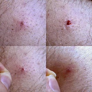
Ingrown hair occurs when the sharp, cut-end of thick hair grows sideways, at an abnormal angle, penetrating into the dermal skin.
Quite often the condition resolves by itself without any clinical symptoms. In some persons ingrown hair may cause small pimple-like bumps with symptoms like reddening, inflammation, pain and pus formation. Though in most of the cases, the ingrowth involves the skin areas which are shaved, tweezed or waxed, it can occur in any hairy part on the body.

Ingrown hair causes

Pseudofolliculitis barbae

Pseudofolliculitis barbae, a type of extensive ingrowth, occurs in persons with thick, coarse hair. It is a common chronic inflammatory disorder in men of African ancestry, occurring most often in regions of thick hair growth after shaving. The condition causes erythematous papules with symptoms of pain and pus formation. A common polymorphism in a keratin gene (K6hf) may be a genetic risk factor for pseudofolliculitis barbae type of ingrowth.

Ingrown hair symptoms

In most of the conditions, the symptoms may be limited to small reddish bumps. The symptoms like rash, tenderness and itching skin may also appear. Infected ingrowth may present symptoms such as pimple like inflammation, raised large bumps, abscesses and pruritic erythematous pustules. Certain other medical conditions like folliculitis, keratosis pilaris and furuncle may mimic the symptoms and are to differentially diagnosed.
image of ingrown hair
Ingrown hair
Common signs and symptoms include:
  • inflammation,
  • papules,
  • pustules,
  • hyperpigmentation,
  • rash,
  • pain,
  • pruritus and
  • embedded hairs in the site.

Ingrown hair treatment

Avoiding shaving for two to three weeks may causes the existing bumps to resolve. Extrafollicular ingrowths can usually be tweezed gently and cut above the skin level. Transfollicular ingrowths as well as ingrowths with symptoms of pus and severe inflammation may require medical treatment. Chemical depilatories, topical creams, laser therapy or surgical removal are the other treatment options available.

Ingrown hair prevention

The best way to prevent the ingrowth is to refrain from shaving or using a beard trimmer. If shaving is necessary, wetting the skin with warm water and using a shaving gel may prevent the hair strand from getting pulled and getting cut close to the root. Avoiding shaving close to skin will enable the cut ends to grow out of the follicle smoothly. Shaving against the direction of the growth of strand must be avoided to prevent ingrown hair.
Advertisements
Related topics in natural skin care:
.
.
.
.
.
.
.
.
.
.
.
.
.
.
.
.
.
.
.
.
.
.
.
.
.
References on ingrown hair:
1.Ribera M, Fernández-Chico N, Casals M. [Pseudofolliculitis barbae]. Actas Dermosifiliogr. 2010 Nov;101(9):749-57.
2.Alexis A, Heath CR, Halder RM. Folliculitis keloidalis nuchae and pseudofolliculitis barbae: are prevention and effective treatment within reach? Dermatol Clin. 2014 Apr;32(2):183-91.
Image source: http://en.wikipedia.org/wiki/File:Eingewachsenes_Haar_2010.jpg
Image author: LBPics | Image license: CC BY-SA 3.0
Interesting topics in natural skin care:
.
.
.
.
.
.
.
.
.
.
.
.
.
.
.
.
.
.
.
.
.
.
.
Current topic in natural skin care: Ingrown hair causes and symptoms

Get glowing skin complexion. Remove acne scars and blemishes from face.

Tuesday, July 21

Chronic paronychia - Fungal (candidal) paronychia

  ›     ›     ›   Chronic fungal and candidal paronychia
What is chronic paronychia?
Chronic paronychia is the persistent or long-lasting inflammatory reaction involving the folds of tissue surrounding a fingernail or toenail.
It is usually caused by the reaction of the proximal nail fold to irritants and allergens and subsequent fungal, more often candidal infection. Candidal paronychia may last for more than six weeks.

Causes of chronic paronychia

Chronic paronychia is prevalent in persons whose hands or feet are frequently exposed to moist conditions. Warm and moist conditions are ideal for contracting fungal infections. Candidal nail fold infection is caused by the fungus species Candida albicans. This multifactorial inflammatory reaction of the nail fold and candidal fungal infection can be as the result some of the causative factors such as,

Signs, symptoms and diagnosis

The nail fold affected by candidal or fungal infection may present with symptoms like reddening (erythema), tenderness and edema (swelling). The cuticle may appear retracted and separated from the nail plate. The proximal nail fold may appear retracted and the adjacent cuticle may be absent. Usually more than one finger is involved and frequently thumb and second or third fingers of the dominant hand are affected by candidal fungal infection. The nail plate gets discolored, thickened and disfigured. Transverse ridges appear due to inflammation and damage to the nail matrix caused by chronic fungal or candidal infection.

The physical examination of the affected nail folds and correlating the symptoms with the history of the presence of causative factors helps in the diagnosis of the nail fold disease. The candidal fungal paronychia may have to be differentiated from other conditions affecting the fingertips, such as squamous cell carcinoma, malignant tumors, eczema and psoriasis.
picture of thumb affected by chronic paronychia and Fungal (candidal) paronychia
Chronic paronychia - Fungal (candidal) paronychia
(Picture author: Rob Hille | CC BY-SA 3.0)

Prevalence and prevention

Chronic fungal nail fold infection and inflammation is prevalent in laundry workers, agricultural laborers, food handlers, drain cleaners, cooks, dishwashers, bartenders, fishermen, fishmongers, fish farm workers, dairy workers, confectioners, nurses, watersports personals and swimmers. Following certain preventive measures can protect a person from developing fungal or candidal infection of the nail fold. Use of gloves, gumboots and avoiding nail fold trauma and irritation can prevent chronic candidal paronychia.

Fungal paronychia treatment

Chronic candidal paronychia is treated by avoiding exposure to the causative factors. In case of milder infection, broad-spectrum topical antifungal cream and a steroid cream is applied. If the infection is severe, oral antifungals and steroids are also prescribed. Recalcitrant candidal or fungal paronychia may be treated by excision of the proximal nail fold or eponychial marsupialization. The resolution of chronic, fungal or candidal paronychia may take several weeks or months and there is the risk of recurrence.
Advertisements
Related topics in natural skin care:
.
.
.
.
.
.
.
.
.
.
.
.
.
.
.
.
.
.
.
.
References:
1.Rigopoulos D, Larios G, Gregoriou S, Alevizos A. Acute and chronic paronychia. Am Fam Physician. 2008 Feb 1;77(3):339-46.
2.Shafritz AB, Coppage JM. Acute and chronic paronychia of the hand. J Am Acad Orthop Surg. 2014 Mar;22(3):165-74.
Interesting topics in natural skin care:
.
.
.
.
.
.
.
.
.
.
.
.
.
.
.
.
.
.
.
.
.
Current topic in natural skin care: Chronic, fungal, candidal paronychia

Get glowing skin complexion. Remove acne scars and blemishes from face.

Thursday, July 2

Acute paronychia - Bacterial (pyogenic) paronychia

  ›     ›     ›   Acute paronychia - Bacterial, pyogenic paronychia
Paronychia is infection and inflammation of the paronychial tissues (nail fold) of the fingers or, less commonly, the toes. Acute paronychia, in most cases, involves bacterial infection which may progress into pyogenic (pus forming) abscess.
Acute paronychia presents as sudden, painful, swollen, red (erythematous), hot, tender nail folds.

The most common infecting organism in bacterial pyogenic paronychia is Staphylococcus aureus. Other Streptococcus species, Pseudomonas species, Gram-negative bacteria and anaerobic bacteria may also be the causative organisms. These pathogens enter the paronychial tissues when there is physical or chemical damage to the nail fold. When treated, acute form of bacterial paronychia usually resolves within 2-3 weeks and usually does not recur. If there is no fluctuance (presence of pus) the infection may resolve with warm soaks. If the infection is pyogenic, oral antibiotic therapy and surgical drainage may help in resolving the condition.
Popular topics:


Causes of acute paronychia

Trauma, physical damage, irritation and chemical damage to the cuticle or nail fold is the main factor associated with the development of pyogenic paronychia. Damage to nail fold may occur from dropping of heavy objects on toes or fingers, manicure procedures, biting or picking at cuticle or hangnail, ingrown nail, dishwashing or any sharp object cutting or piercing into nail fold tissue. Such damage to the paronychial tissues or cuticle allows the entry of pyogenic pathogens.

Certain habits like nail biting, biting or picking at a hangnail or finger sucking may also give rise to pyogenic bacterial infection of the paronychial tissues. Exposure to saliva and oral flora, will facilitate the entry of several anaerobic gram-negative pyogenic bacteria into paronychial tissues.

The most common infecting organism in this acute pyogenic infection is Staphylococcus aureus. Other causative organisms include Streptococcus pyogenes, Pseudomonas pyocyanea, Proteus vulgaris, other Streptococcus species, Pseudomonas species, Gram-negative bacteria and anaerobic bacteria.

Signs, symptoms and clinical manifestations

In acute pyogenic paronychia, typically only one nail is involved. The signs include reddening (erythema) and swelling (edema) of the proximal and lateral nail folds. Initially, it presents as superficial bacterial infection. Later it becomes pyogenic with accumulation of purulent material under the nail fold. The symptoms include extreme pain, pressure, discomfort and tenderness. The patient gets great relief when the pus is drained. An untreated acute paronychia may progress to infect the nail matrix leading to permanent dystrophy of the nail plate.
picture of acute, bacterial, pyogenic paronychia

Diagnosis of acute paronychia

The history of recent minor trauma and physical examination of nail folds will help in the diagnosis of acute paronychia. In early stages, the digital pressure test may be helpful in determining the presence or extent of an pyogenic abscess. The affected area gets blanched and helps in clear demarcation of the abscess. In bacterial paronychia not responding to antibiotics, methicillin-resistant S. aureus (MRSA) infection should be ruled out. Psoriasis, reactive arthritis, dactylitis and herpetic whitlow may have to be differentiated from acute pyogenic paronychia.

Bacterial paronychia treatment

Mild paronychia can be treated at home by soaking the infected nail in warm water or dilute vinegar solution for 3-4 times a day. Acetaminophen or a nonsteroidal anti-inflammatory medication may be taken orally to provide relief from pain and inflammation. A combination of topical antibiotic and corticosteroid cream may be applied. The bacterial infection may heal on its own in a few days.

If the paronychia persists after a week or becomes acute and pyogenic, it requires medical intervention. Persisting infection can be treated by oral antibiotic therapy and surgical drainage. A broad-spectrum oral antibiotic may be prescribed.

To drain the pus, the nail fold is lifted with a 23-gauge needle. This helps in passive draining of the accumulated pus. Mild pressure may be applied on the infected area to facilitate draining. Alternatively a small incision may be made with a scalpel to open up the paronychia abscess and clean it of pus. In severe acute bacterial pyogenic paronychia, the infection may involve nail bed and need partial or complete nail removal.
Advertisements
Related topics in natural skin care:
.
.
.
.
.
.
.
.
.
.
.
.
.
.
.
.
.
.
.
.
References for acute bacterial pyogenic paronychia:
1.Rigopoulos D, Larios G, Gregoriou S, Alevizos A. Acute and chronic paronychia. Am Fam Physician. 2008 Feb 1;77(3):339-46.
2.Shafritz AB, Coppage JM. Acute and chronic paronychia of the hand. J Am Acad Orthop Surg. 2014 Mar;22(3):165-74.
Interesting topics in natural skin care:
.
.
.
.
.
.
.
.
.
.
.
.
.
.
.
.
.
.
.
.
Current topic in natural skin care: Acute, bacterial and pyogenic paronychia.

Get glowing skin complexion. Remove acne scars and blemishes from face.